| Back to "Normal samples" |
Normal Skeletal Muscle - Gomori Trichrome
Last updated on Monday, April 20 2009 by gliageek
|
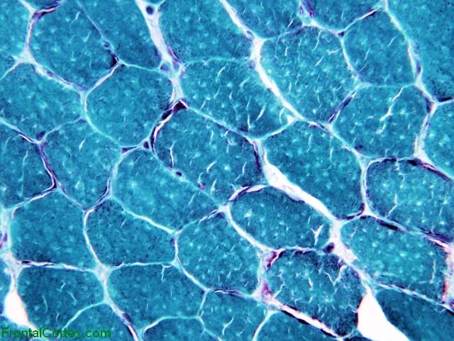
| This is a sample of normal muscle prepared with the Gomori Trichrome stain. The muscle fibers appear a bluish green. Note that normal muscle fibers have a polygonal shape to them, and are not exceptionally rounded. Note also that the muscle fibers tend to be about the same size. The nuclei (which appear dark) are located laterally, near the cell membrane. There are small white cracks in each muscle fiber - these are an artifact caused during preparation of the sample. Another common artifact (not shown) is patches of bright red staining secondary to partial fixation of the muscle due to exposure to formalin fumes. |
Categories (tags) users associate with this resource 
Please type in an appropriate tag for this item
Click on a tag to find related images, videos, MCQs, and other resources.
Check the boxes next to the tags you consider relevant or enter your own tags in the field below.
You must be logged in to edit tags.
Top 5 tags for this item:
No tags have been created yet for this resource.Please type in an appropriate tag for this item
more tags:
new tag:
log in to FrontalCortex.com
New to FrontalCortex?
|

![]()
![]()

| | We comply with the HONcode standard for trustworthy health information: verify here. |
Share this page:
 |  |
 |
 |
 |
 |
 |
 |
 |
 |
 |
 |
 |
|
Wednesday, April 24, 2024 at 11:03:33 PM
This site has been visited 45849652 times since June 6 2006
All software and content (C) 2004-2024, FrontalCortex, Inc. unless otherwise specified.
privacy policy Web 2.0 policy disclaimer contact us
All software and content (C) 2004-2024, FrontalCortex, Inc. unless otherwise specified.
privacy policy Web 2.0 policy disclaimer contact us