| Back to "Acute neuronal necrosis - cerebellum" |
Acute neuronal necrosis cerebellum 2
Last updated on Tuesday, September 2 2008 by jdmiles
|
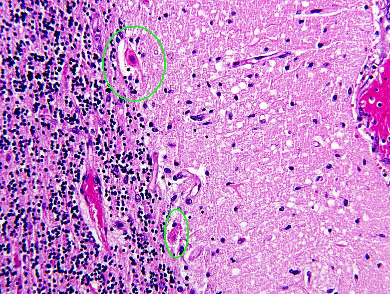
| The cells circled in green in this image are Purkinje cells. Purkinje cells are found in the cerebellum between the granule cell layer (dark blue cells on the left) and the molecular layer (the big pink area taking up most of the right side of this picture). These Purkinje cells are bright red, which is an indicator of injury. Purkinje cells are very sensitive to hypoxia, and are among the first cells to die in cerebral ischemia. Note that it is only the Purkinje cells which are bright red - the granule cells and the cells in the molecular layer remain blue. |
Categories (tags) users associate with this resource 
Please type in an appropriate tag for this item
Click on a tag to find related images, videos, MCQs, and other resources.
Check the boxes next to the tags you consider relevant or enter your own tags in the field below.
You must be logged in to edit tags.
Top 5 tags for this item:
No tags have been created yet for this resource.Please type in an appropriate tag for this item
more tags:
new tag:
log in to FrontalCortex.com
New to FrontalCortex?
|

![]()
![]()

| | We comply with the HONcode standard for trustworthy health information: verify here. |
Share this page:
 |  |
 |
 |
 |
 |
 |
 |
 |
 |
 |
 |
 |
|
Friday, April 19, 2024 at 6:58:42 PM
This site has been visited 45819044 times since June 6 2006
All software and content (C) 2004-2024, FrontalCortex, Inc. unless otherwise specified.
privacy policy Web 2.0 policy disclaimer contact us
All software and content (C) 2004-2024, FrontalCortex, Inc. unless otherwise specified.
privacy policy Web 2.0 policy disclaimer contact us