| Back to "Leukodystrophies" |
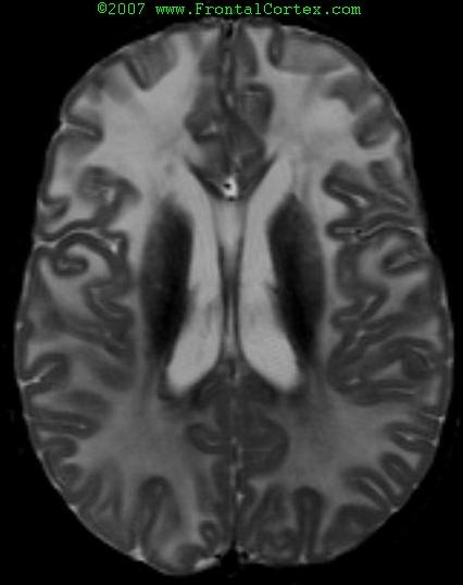
Alexander Disease on T2-weighted MRI
Last updated on Sunday, March 29 2009 by jdmiles
|
| The findings in this MRI are typical for Alexander disease. MRI criteria for the diagnosis of Alexander disease include: 1. Extensive, frontally-predominant cerebral white matter changes 2. A periventricular rim which is bright on T1 and dark on T2 3. Brainstem abnormalities 4. Abnormalities of hte basal ganglia and thalami 5. Contrast enhancement of particular gray and white matter structures. Four of 5 criteria should be met to make the diagnosis based on imaging. |
Categories (tags) users associate with this resource 
Please type in an appropriate tag for this item
Click on a tag to find related images, videos, MCQs, and other resources.
Check the boxes next to the tags you consider relevant or enter your own tags in the field below.
You must be logged in to edit tags.
Top 5 tags for this item:
No tags have been created yet for this resource.Please type in an appropriate tag for this item
more tags:
new tag:
log in to FrontalCortex.com
New to FrontalCortex?
|

![]()
![]()

| | We comply with the HONcode standard for trustworthy health information: verify here. |
Share this page:
 |  |
 |
 |
 |
 |
 |
 |
 |
 |
 |
 |
 |
|
Thursday, April 18, 2024 at 11:35:44 AM
This site has been visited 45812071 times since June 6 2006
All software and content (C) 2004-2024, FrontalCortex, Inc. unless otherwise specified.
privacy policy Web 2.0 policy disclaimer contact us
All software and content (C) 2004-2024, FrontalCortex, Inc. unless otherwise specified.
privacy policy Web 2.0 policy disclaimer contact us