| Back to "COX (Cytochrome Oxidase) Staining" |
Cytochrome Oxidase staining - Example of pathological findings
Last updated on Saturday, April 4 2009 by jdmiles
|
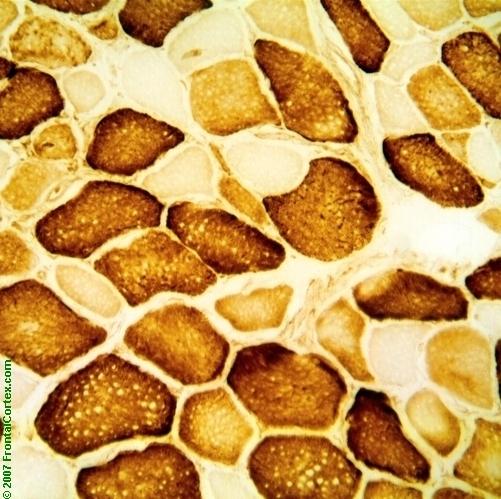
| This is a cytochrome oxidase c stain (cox stain) of a skeletal muscle biopsy. Cytochrome C oxidase is a marker of mitochondrial activity. Type I muscle fibers have a higher capacity for oxidative metabolism than type II cells, which have a higher capacity for anaerobic metabolism. This means that the type I muscle cells will stain darker on cox, but both populations should show some uptake of the brown pigment. Normally, the 2 muscle groups represent the only 2 populations seen on cox staining. However, this muscle is affected by a mitochondrial myopathy, and shows a third population: occasional cox-negative fibers, which appear very pale, indicating almost no oxidative metabolism. |
Related images:
|
| Cytochrome Oxidase staining - Example of pathological findings with arrow The unnecessarily large green arrow points to a pale, cytochrome oxidase negative fiber. This fiber has little or no capacity for oxidative metabolism. |
Categories (tags) users associate with this resource 
Please type in an appropriate tag for this item
Click on a tag to find related images, videos, MCQs, and other resources.
Check the boxes next to the tags you consider relevant or enter your own tags in the field below.
You must be logged in to edit tags.
Top 5 tags for this item:
No tags have been created yet for this resource.Please type in an appropriate tag for this item
more tags:
new tag:
log in to FrontalCortex.com
New to FrontalCortex?
|

![]()
![]()

| | We comply with the HONcode standard for trustworthy health information: verify here. |
Share this page:
 |  |
 |
 |
 |
 |
 |
 |
 |
 |
 |
 |
 |
|
Friday, April 19, 2024 at 8:30:58 AM
This site has been visited 45816483 times since June 6 2006
All software and content (C) 2004-2024, FrontalCortex, Inc. unless otherwise specified.
privacy policy Web 2.0 policy disclaimer contact us
All software and content (C) 2004-2024, FrontalCortex, Inc. unless otherwise specified.
privacy policy Web 2.0 policy disclaimer contact us