This image shows:
|
| |||||||||||||||||||||||||||||||||||||||||||||||||||||||||||||||||||||||||||||||||||||||||||||||||||||||
Cerebrovascular Anatomy 1Topic: AnatomyCreated on Friday, August 24 2007 by jdmiles Last modified on Friday, August 24 2007.
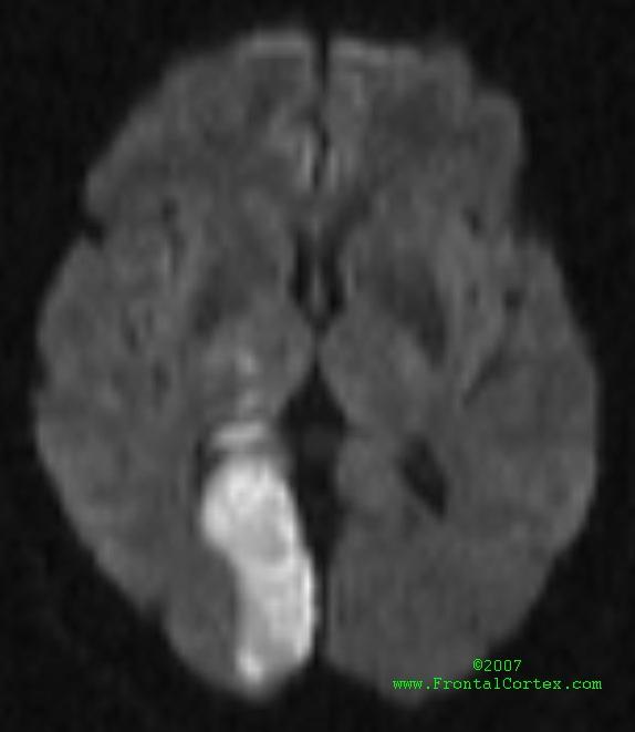
This image shows: A) MRI diffusion-weighted image of a stroke in the territory of left PICA B) MRI diffusion-weighted image of a stroke in the territory of right MCA C) MRI diffusion-weighted image of a stroke in the territory of left SCA D) MRI diffusion-weighted image of a stroke in the territory of left MCA E) MRI diffusion-weighted image of a stroke in the territory of right PCA
This question was last modified on August 24, 2007.
ANSWERS AND EXPLANATIONSA) MRI diffusion-weighted image of a stroke in the territory of left PICA
| |||||||||||||||||||||||||||||||||||||||||||||||||||||||||||||||||||||||||||||||||||||||||||||||||||||||
 |  |  | 
|  |  |
| Please log in if you want to rate questions. | |||||
This is an MRI diffusion weighted image. The bright area represents diffusion restriction, which can be seen in acute stroke. The area of diffusion restriction corresponds to the right posterior cerebral artery (PCA) territory.
The middle cerebral artery (MCA) does not supply the territory where this stroke occurred.
(See References) | | |
| | |
| Please log in if you want to rate questions. | |||||
The superior cerebellar artery (SCA) does not supply the territory where this stroke occurred.
(See References) | | |
| | |
| Please log in if you want to rate questions. | |||||
The middle cerebral artery (MCA) does not supply the territory where this stroke occurred.
(See References) | | |
| | |
| Please log in if you want to rate questions. | |||||
| | |
| | |
| Please log in if you want to rate questions. | |||||
| 1. Nolte, J. (1993). The Human Brain: An Introduction to Its Functional Anatomy. Mosby, St. Louis. |
| | |
| | |
| Please log in if you want to rate questions. | |||||
FrontalCortex.com -- Neurology Review Questions -- Neurology Boards -- Board Review -- Residency Inservice Training Exam -- RITE Exam Review

![]()
![]()

| | We comply with the HONcode standard for trustworthy health information: verify here. |
Share this page:
 |  |
 |
 |
 |
 |
 |
 |
 |
 |
 |
 |
 |
|