| Back to "Alexander disease, coronal section, close-up view" |
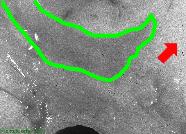
Alexander disease, coronal section, close-up view, highlighted
Last updated on Sunday, March 29 2009 by jdmiles
|
| This is a slice of brain from a patient who had Alexander disease. In this disease, there is diffuse loss of white matter. In this image, note how dark the demyelinated white matter appears, especially in contrast to the relatively spared region indicated by the unnecessarily large red arrow. Note also that the subcortical U-fibers (outlined in green) are also demyelinated. This finding can be of diagnostic value when trying to differentiate different leukodystrophies. |
Categories (tags) users associate with this resource 
Please type in an appropriate tag for this item
Click on a tag to find related images, videos, MCQs, and other resources.
Check the boxes next to the tags you consider relevant or enter your own tags in the field below.
You must be logged in to edit tags.
Top 5 tags for this item:
No tags have been created yet for this resource.Please type in an appropriate tag for this item
more tags:
new tag:
log in to FrontalCortex.com
New to FrontalCortex?
|

![]()
![]()

| | We comply with the HONcode standard for trustworthy health information: verify here. |
Share this page:
 |  |
 |
 |
 |
 |
 |
 |
 |
 |
 |
 |
 |
|
Wednesday, April 01, 2026 at 4:47:52 AM
This site has been visited 52424005 times since June 6 2006
All software and content (C) 2004-2026, FrontalCortex, Inc. unless otherwise specified.
privacy policy Web 2.0 policy disclaimer contact us
All software and content (C) 2004-2026, FrontalCortex, Inc. unless otherwise specified.
privacy policy Web 2.0 policy disclaimer contact us