| Back to "Dorsolateral Medullary Infarct, transverse section." |
Wallenberg Syndrome MRI
Last updated on Saturday, April 11 2009 by jdmiles
|
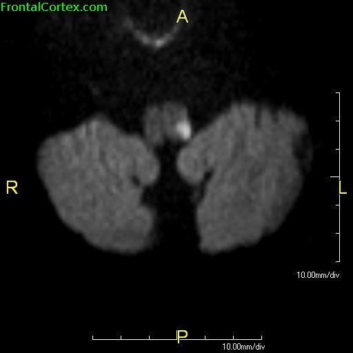
| Diffusion-weighted MRI of an infart in the left dorsolateral medulla, consistent with a clinical picture of Wallenberg syndrome.
The infarct in this image would be in approximately the same location as the one shown in the pathology image. In the MRI, dorsal is toward the bottom of the image, whereas in the pathology image, dorsal is toward the top. Also, in MRI imaging, anatomic left is on the right of the image, whereas in photographs of pathology specimens, it is customary for anatomic left to be on the left. Courtesy of Wikimedia. Image created by John S. To, M.D. This image is in the public domain. |
Related images:
|
| Wallenberg Syndrome MRI with arrow Diffusion-weighted MRI of an infart in the left dorsolateral medulla, consistent with a clinical picture of Wallenberg syndrome. The infarct in this image would be in approximately the same location as the one shown in the pathology image. In the MRI, dorsal... |
Categories (tags) users associate with this resource 
Please type in an appropriate tag for this item
Click on a tag to find related images, videos, MCQs, and other resources.
Check the boxes next to the tags you consider relevant or enter your own tags in the field below.
You must be logged in to edit tags.
Top 5 tags for this item:
No tags have been created yet for this resource.Please type in an appropriate tag for this item
more tags:
new tag:
log in to FrontalCortex.com
New to FrontalCortex?
|

![]()
![]()

| | We comply with the HONcode standard for trustworthy health information: verify here. |
Share this page:
 |  |
 |
 |
 |
 |
 |
 |
 |
 |
 |
 |
 |
|
Tuesday, July 01, 2025 at 8:38:29 PM
This site has been visited 49821680 times since June 6 2006
All software and content (C) 2004-2025, FrontalCortex, Inc. unless otherwise specified.
privacy policy Web 2.0 policy disclaimer contact us
All software and content (C) 2004-2025, FrontalCortex, Inc. unless otherwise specified.
privacy policy Web 2.0 policy disclaimer contact us