| Back to "Vascular Disease 1: Reaction to ischemic injury" |
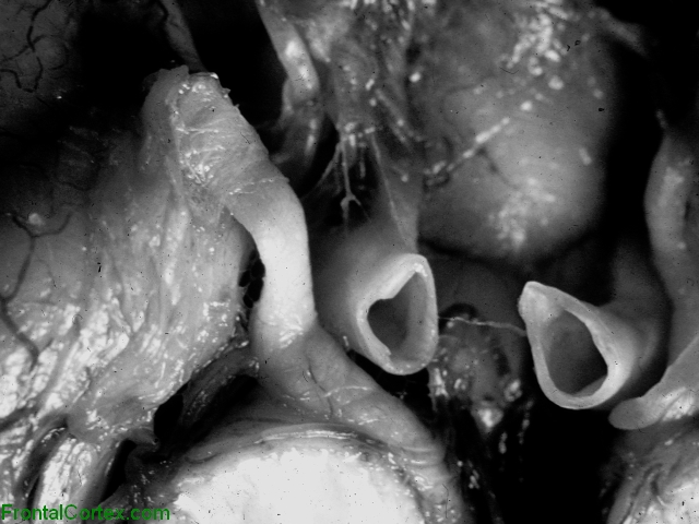
Transtentorial herniation, close up view of ventral surface of brain.
Last updated on Wednesday, April 15 2009 by gliageek
|
| This close up photograph demonstrates compression of the oculomotor nerve against the (removed) tentorium cerebelli by the expanding hemispheric brain parenchyma. As the parasympathetic innervation to the anterior eye is carried by fibers on the surface of this nerve, compression results in unopposed sympathetic activity to the ciliary muscle, manifested as a fixed, dilated pupil. |
Categories (tags) users associate with this resource 
Please type in an appropriate tag for this item
Click on a tag to find related images, videos, MCQs, and other resources.
Check the boxes next to the tags you consider relevant or enter your own tags in the field below.
You must be logged in to edit tags.
Top 5 tags for this item:
No tags have been created yet for this resource.Please type in an appropriate tag for this item
more tags:
new tag:
log in to FrontalCortex.com
New to FrontalCortex?
|

![]()
![]()

| | We comply with the HONcode standard for trustworthy health information: verify here. |
Share this page:
 |  |
 |
 |
 |
 |
 |
 |
 |
 |
 |
 |
 |
|
Thursday, April 25, 2024 at 11:07:30 PM
This site has been visited 45855042 times since June 6 2006
All software and content (C) 2004-2024, FrontalCortex, Inc. unless otherwise specified.
privacy policy Web 2.0 policy disclaimer contact us
All software and content (C) 2004-2024, FrontalCortex, Inc. unless otherwise specified.
privacy policy Web 2.0 policy disclaimer contact us