| Back to "Miscellaneous Demyelinating Diseases" |
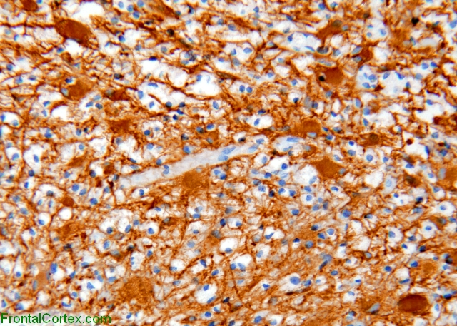
Tumefactive demyelination, immunohistochemical staining for glial fibrillary acidic protein
Last updated on Thursday, March 26 2009 by gliageek
|
| GFAP immunohistochemical staining demonstrates evenly spaced reactive astrocytes with abundant processes and relatively small eccentric nuclei. Macrophages stand out as negatively stained cells within the fibrillary brown background. |
Categories (tags) users associate with this resource 
Please type in an appropriate tag for this item
Click on a tag to find related images, videos, MCQs, and other resources.
Check the boxes next to the tags you consider relevant or enter your own tags in the field below.
You must be logged in to edit tags.
Top 5 tags for this item:
No tags have been created yet for this resource.Please type in an appropriate tag for this item
more tags:
new tag:
log in to FrontalCortex.com
New to FrontalCortex?
|

![]()
![]()

| | We comply with the HONcode standard for trustworthy health information: verify here. |
Share this page:
 |  |
 |
 |
 |
 |
 |
 |
 |
 |
 |
 |
 |
|
Friday, April 26, 2024 at 8:04:18 PM
This site has been visited 45863767 times since June 6 2006
All software and content (C) 2004-2024, FrontalCortex, Inc. unless otherwise specified.
privacy policy Web 2.0 policy disclaimer contact us
All software and content (C) 2004-2024, FrontalCortex, Inc. unless otherwise specified.
privacy policy Web 2.0 policy disclaimer contact us