Neuromuscular 10
Topic: Pathology
Created on Thursday, February 26 2009 by jdmiles
Last modified on Thursday, February 26 2009.
Courtesy of Dr. Mark Cohen
What does this biopsy specimen show?
This question was created on February 26, 2009 by jdmiles.
This question was last modified on February 26, 2009.
ANSWERS AND EXPLANATIONS
A) A Meningioma
This answer is incorrect.
No. This is muscle. (
See References)
|
 |  |  | 
|  |  |
| Please log in if you want to rate questions. |
B) Muscle from someone with really bad neuropathy
This answer is incorrect.
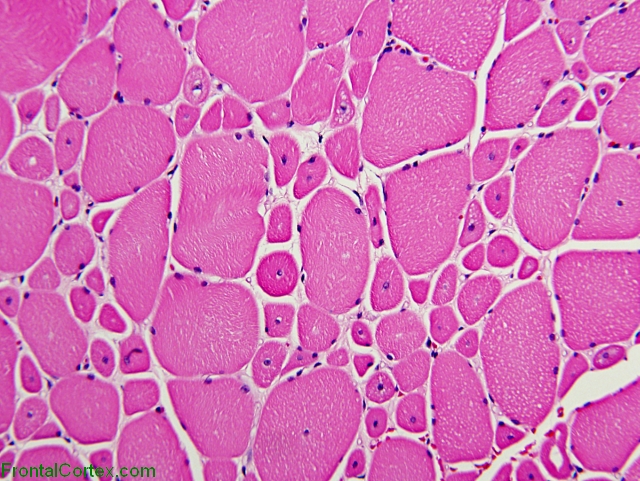
The image in question (above, at the top of this page) is an H&E-stained biopsy of skeletal muscle from a person with centronuclear myopathy.
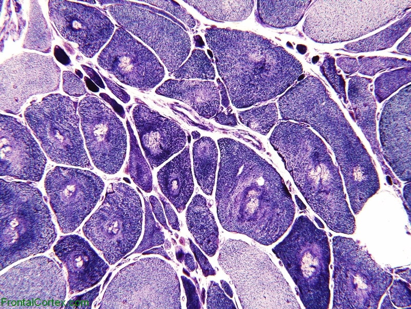
The image below shows an NADH-stained muscle biopsy from someone with denervated (i.e., neuropathic) muscle. A couple of classic neuropathic changes include increased angularity of muscle fibers, formation of target fibers. both of these features can be seen in the image below, but not in the image at the top of this page, which the question asked about.
Courtesy of Dr. Mark Cohen
Please note that target fibers can be easily confused with central cores -- they are not the same thing. Use the search bar to look for "CCD" or "Core" or "Central core disease" for more specific information.
(
See References)
|
| | |
| | |
| Please log in if you want to rate questions. |
C) Myopathic muscle
This answer is correct.
This is an H&E-stained biopsy of skeletal muscle from a person with centronuclear myopathy.
Classic myopathic changes are seen in this image: fiber size variability, rounding of muscle fibers, and centrally located nuclei.
Fiber Type Variability
The muscle fibers are varied in size - some are quite large, some are very small. In a biopsy of normal muscle, most muscle fibers would appear to be about the same size.
Rounded Fibers
Normally, muscle fibers cut in cross-section have a polygonal appearance. In neuropathy, the angulation of the fibers becomes exaggerated. In myopathy, the fibers become more rounded.
Centrally Located Nuclei
Muscle fibers are polynuclear syncytia rather than cells with a single nucleus. The nuclei in normal muscle fibers are located on the periphery, close to the cell membrane. In myopathy, the muscle fibers get more centrally located.
For more information, check out gliageek's video presentation on "Denervation, Dystrophy, and Disorders of Energy Metabolism" (search for it in the search bar, or check out the link at the bottom of this page). (
See References)
|
| | |
| | |
| Please log in if you want to rate questions. |
D) A Bear.
This answer is incorrect.
No. The image at the top of the page is muscle.
Bears look like this:
(
See References)
|
| | |
| | |
| Please log in if you want to rate questions. |
E) Normal muscle
This answer is incorrect.
The image in question (above, at the top of this page) is an H&E-stained biopsy of skeletal muscle from a person with centronuclear myopathy.
Normal muscle would not be as rounded. The fibers in normal muscle would not have as much variation in size. A sample of normal muscle is shown below:
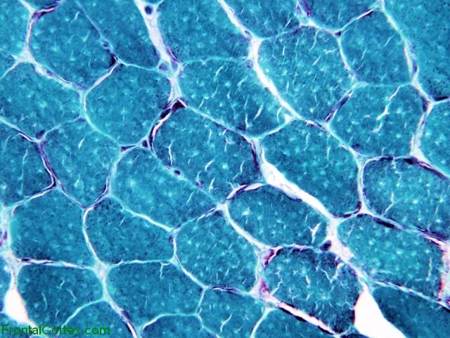
Courtesy of Dr. Mark Cohen
(
See References)
|
| | |
| | |
| Please log in if you want to rate questions. |
References:
| 1. Prayson, R.A., and Goldblum, J.R. (Eds.) (2005). Neuropathology. Elsevier Churchill Livingstone, Philadelphia. (ISBN:0443066582) | Advertising:
|
|
| | |
| | |
| Please log in if you want to rate questions. |
FrontalCortex.com -- Neurology Review Questions -- Neurology Boards -- Board Review -- Residency Inservice Training Exam -- RITE Exam Review
pathology
Neuromuscular 10
Question ID: 022609204
Question written by J. Douglas Miles, (C) 2006-2009, all rights reserved.
Created: 02/26/2009
Modified: 02/26/2009
Estimated Permutations: 120