Basic Neuropathology 7
Topic: Imaging
Created on Thursday, February 14 2013 by gliageek
Last modified on Thursday, February 14 2013.
A) Herpes simplex encephalitis
B) Frontotemporal lobar degeneration
C) Human immunodeficiency virus associated dementia
D) Limbic encephalitis
E) Jakob-Creutzfeldt disease
 | = Go back to the top of the page. |
 | = Reload a different version of this question (). |
 | = Load a random question from the database. |

| = Use this question as a template to create a totally NEW question. |
 | = Enter detailed rating for this question! |
Average rating not yet available | = How users like you have rated this question. |
This question was last modified on February 14, 2013.
ANSWERS AND EXPLANATIONS
A) Herpes simplex encephalitis
This answer is incorrect.
Herpes simplex encephalitis is characterized by severe necrosis in addition to the findings seen within this slide. (See References)
| | |
| | Average rating not yet available |
| Please log in if you want to rate questions. | |||||
B) Frontotemporal lobar degeneration
This answer is incorrect.
Frontotemporal lobar degeneration is a noninflammatory degenerative condition, and does not show microglia nodules. (See References)
| | |
| | Average rating not yet available |
| Please log in if you want to rate questions. | |||||
C) Human immunodeficiency virus associated dementia
This answer is incorrect.
Although microglial nodules are seen within HAD, they're located predominantly within the white matter and basal ganglia, and usually demonstrate the presence of multi-nucleated microglial cells resulting from viral induced fusion. (See References)
| | |
| | Average rating not yet available |
| Please log in if you want to rate questions. | |||||
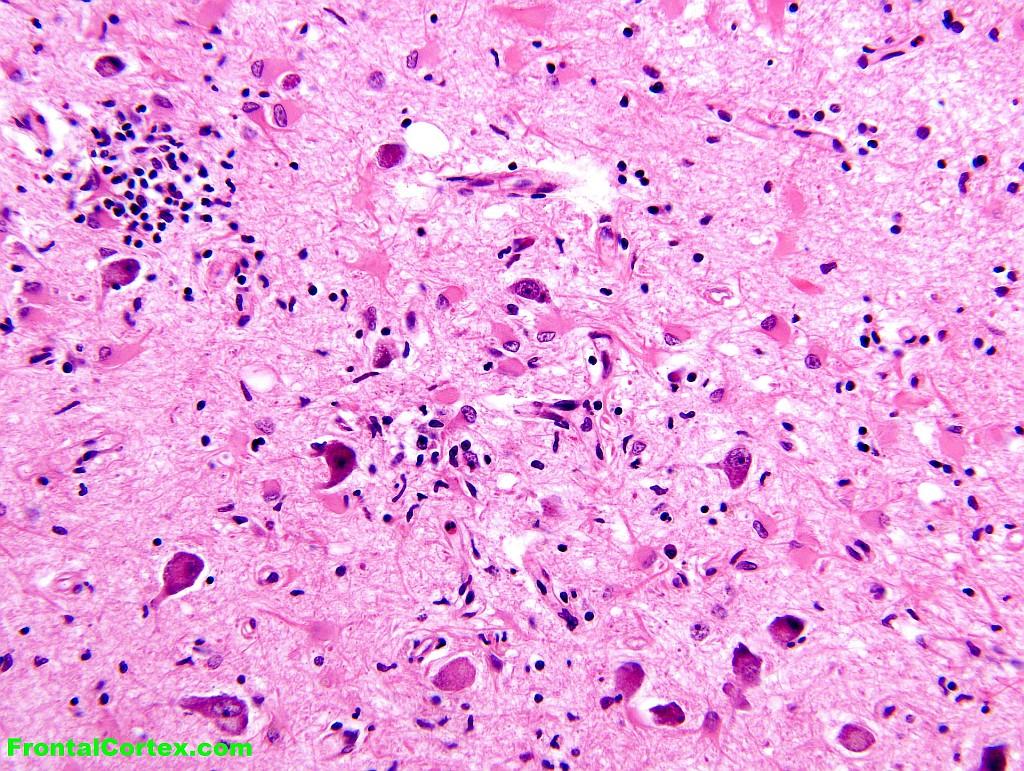
D) Limbic encephalitis
This answer is correct.
The presence of microglia nodules with brisk reactive astrocytosis without liquefactive necrosis characterizes limbic encephalitis. this is characteristically, but not invariably, associated with underlying malignancy (specifically, small cell carcinoma of the lung). (See References)
| | |
| | Average rating not yet available |
| Please log in if you want to rate questions. | |||||
E) Jakob-Creutzfeldt disease
This answer is incorrect.
Prion diseases, such as Jakob-Creutzfeldt disease, are characterized by the absence of an inflammatory infiltrate, as the pathogenic protein is a confirmational variant of a normal cellular protein, and is hence not recognized as foreign body immune system. (See References)
| | |
| | Average rating not yet available |
| Please log in if you want to rate questions. | |||||
References:
| | |
| | Average rating not yet available |
| Please log in if you want to rate questions. | |||||
FrontalCortex.com -- Neurology Review Questions -- Neurology Boards -- Board Review -- Residency Inservice Training Exam -- RITE Exam Review
imaging
Basic Neuropathology 7
Question ID: 021413084
Question written by gliageek. (C) FrontalCortex.com 2006-2009, all rights reserved. Created: 02/14/2013
Modified: 02/14/2013
Estimated Permutations: 120
User Comments About This Question:
log in to FrontalCortex.com
New to FrontalCortex?
|

![]()
![]()

| | We comply with the HONcode standard for trustworthy health information: verify here. |
Share this page:
 |  |
 |
 |
 |
 |
 |
 |
 |
 |
 |
 |
 |
|
Saturday, July 05, 2025 at 7:51:52 PM
This site has been visited 49851758 times since June 6 2006
All software and content (C) 2004-2025, FrontalCortex, Inc. unless otherwise specified.
privacy policy Web 2.0 policy disclaimer contact us
All software and content (C) 2004-2025, FrontalCortex, Inc. unless otherwise specified.
privacy policy Web 2.0 policy disclaimer contact us