| Back to "Glioblastoma multiforme involving cerebellum, H&E stain x 100" |
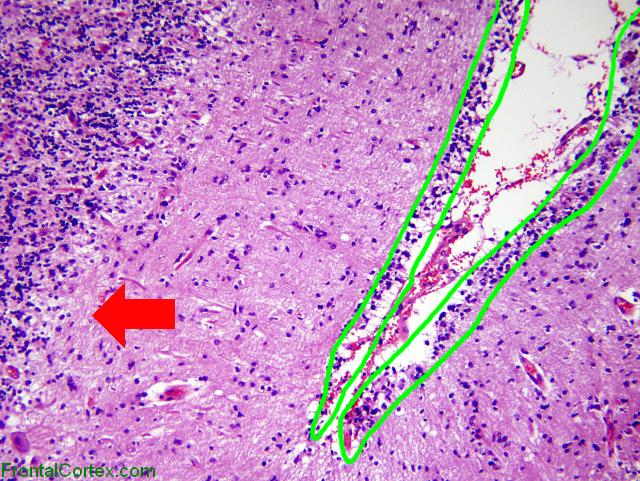
Glioblastoma multiforme involving cerebellum, H&E stain x 100, Labeled
Last updated on Saturday, April 4 2009 by jdmiles
|
| This slide shows spread of a glioblastoma to the cerebellum. The diagnosis of GBM is not clearly evident from this slide, but this is from the same patient as the slide showing the necrosis, so the diagnosis is known. The most significant finding in this image is the area highlighted in green - what looks like infant external granular cell layer is actually subpial spread of tumor cells. This person was not an infant. It is also interesting to note that there is a paucity of Purkinje cells in the Purkinje cell layer (unnecessarily large red arrow). |
Categories (tags) users associate with this resource 
Please type in an appropriate tag for this item
Click on a tag to find related images, videos, MCQs, and other resources.
Check the boxes next to the tags you consider relevant or enter your own tags in the field below.
You must be logged in to edit tags.
Top 5 tags for this item:
No tags have been created yet for this resource.Please type in an appropriate tag for this item
more tags:
new tag:
log in to FrontalCortex.com
New to FrontalCortex?
|

![]()
![]()

| | We comply with the HONcode standard for trustworthy health information: verify here. |
Share this page:
 |  |
 |
 |
 |
 |
 |
 |
 |
 |
 |
 |
 |
|
Wednesday, May 01, 2024 at 11:02:43 PM
This site has been visited 45901106 times since June 6 2006
All software and content (C) 2004-2024, FrontalCortex, Inc. unless otherwise specified.
privacy policy Web 2.0 policy disclaimer contact us
All software and content (C) 2004-2024, FrontalCortex, Inc. unless otherwise specified.
privacy policy Web 2.0 policy disclaimer contact us