| Back to "Commonly used stains" |
Example of immune staining
Last updated on Thursday, August 28 2008 by jdmiles
|
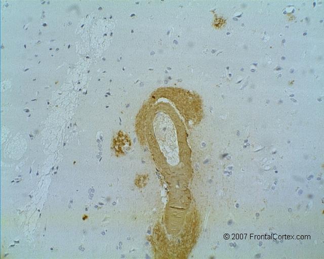
| This is an example of a typical immune stain. The tissue we're looking at is brain, and the stain is an immune stain specific for beta amyloid. In this case, the background is kind of a homogenous gray. The only stuff we're interested in seeing here is brown - this is amyloid. In this slice of brain, we can see brown blobs in the neuropil, which represent senile plaques. We can also see amyloid that has collected in the wall of the blood vessel (the big thing in the middle), which occurs in amyloid angiopathy. Note that we cannot tell just from the colors of this slide which immune stain we're using. |
Related images:
|
| Amyloid Angiopathy Labeled A photomicrograph of a slice of brain tissue, which is stained with an immune stain that is specific for amyloid. The unnecessarily large red arrows in this image point to amyloid plaques and amyloid in a blood vessel. |
|
| Temporary Image This is a test |
Categories (tags) users associate with this resource 
Please type in an appropriate tag for this item
Click on a tag to find related images, videos, MCQs, and other resources.
Check the boxes next to the tags you consider relevant or enter your own tags in the field below.
You must be logged in to edit tags.
Top 5 tags for this item:
No tags have been created yet for this resource.Please type in an appropriate tag for this item
more tags:
new tag:
log in to FrontalCortex.com
New to FrontalCortex?
|

![]()
![]()

| | We comply with the HONcode standard for trustworthy health information: verify here. |
Share this page:
 |  |
 |
 |
 |
 |
 |
 |
 |
 |
 |
 |
 |
|
Friday, April 26, 2024 at 8:14:05 AM
This site has been visited 45859572 times since June 6 2006
All software and content (C) 2004-2024, FrontalCortex, Inc. unless otherwise specified.
privacy policy Web 2.0 policy disclaimer contact us
All software and content (C) 2004-2024, FrontalCortex, Inc. unless otherwise specified.
privacy policy Web 2.0 policy disclaimer contact us