Have you seen the parietal watch?

Neuromuscular 06

Topic: Pathology

Created on Saturday, February 14 2009 by gliageek

Last modified on Saturday, February 14 2009.


Courtesy of Dr. Mark Cohen
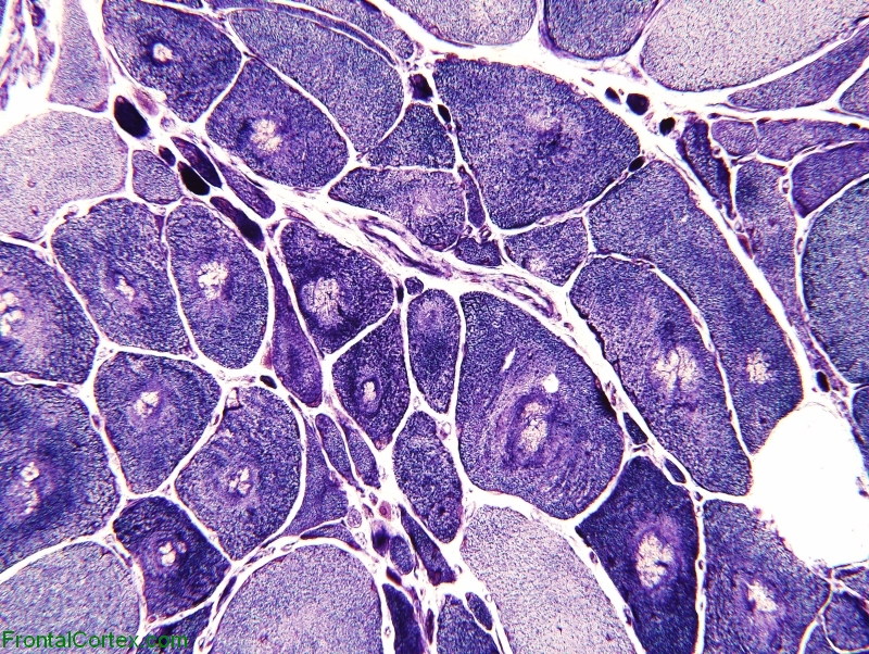
This NADH-TR stained muscle shows features most consistent with:

 
        A) Inclusion bodies
 
        B) Plaquenil myopathy
 
        C) Acid maltase deficiency
 
        D) Reinnervation
 
        E) Rhabdomyolysis


Back to the question = Go back to the top of the page.
See another question like this one = Reload a different version of this question ().
Click here for a random question = Load a random question from the database.
Clone this question = Use this question as a template to create a totally NEW question.
Rate this question = Enter detailed rating for this question!
Average user rating for this question = 5 = How users like you have rated this question.
This question was created on February 14, 2009 by gliageek.
This question was last modified on February 14, 2009.


ANSWERS AND EXPLANATIONS


A) Inclusion bodies

This answer is incorrect.


These show central clearing with surrounding granular material, best seen on H&E and trichrome  (See References)

Back to the questionSee another question like this oneClick here for a random questionClone this question Rate this questionAverage user rating for this question = 5
Please log in if you want to rate questions.


B) Plaquenil myopathy

This answer is incorrect.


Choloroquine & hydroxychloroquine myopathy demonstrates multiple smaller vaculoles within muscle fibers, representing dilated lysosomes  (See References)

Back to the questionSee another question like this oneClick here for a random questionClone this question Rate this questionAverage user rating for this question = 5
Please log in if you want to rate questions.


C) Acid maltase deficiency

This answer is incorrect.


Lysosomal glycogen storage does not show peripheral increase in NADH-TR staining  (See References)

Back to the questionSee another question like this oneClick here for a random questionClone this question Rate this questionAverage user rating for this question = 5
Please log in if you want to rate questions.


D) Reinnervation

This answer is correct.


As atrophied fibers replenish contractile elements after reinnervated, central areas are initially devoid of mitochondria, sarcoplasmic reticulin and ATPase, known as target fibers  (See References)

Back to the questionSee another question like this oneClick here for a random questionClone this question Rate this questionAverage user rating for this question = 5
Please log in if you want to rate questions.


E) Rhabdomyolysis

This answer is incorrect.


Moth-eaten fibers may be seen in some cases of rhabdomyolysis  (See References)

Back to the questionSee another question like this oneClick here for a random questionClone this question Rate this questionAverage user rating for this question = 5
Please log in if you want to rate questions.


References:

1. Graham, D.I., and Lantos, P.L. (2002). Greenfield's Neuropathology, 7th ed. Arnold Press, New York. (ISBN:0340742313)Advertising:
Back to the questionSee another question like this oneClick here for a random questionClone this question Rate this questionAverage user rating for this question = 5
Please log in if you want to rate questions.


FrontalCortex.com -- Neurology Review Questions -- Neurology Boards -- Board Review -- Residency Inservice Training Exam -- RITE Exam Review
pathology
Neuromuscular 06
Question ID: 021409127
Question written by gliageek. (C) FrontalCortex.com 2006-2009, all rights reserved. Created: 02/14/2009
Modified: 02/14/2009
Estimated Permutations: 120

User Comments About This Question:

0 user entries
Please log in if you'd like to add a comment.