Pediatric Epilepsy Syndromes 11
Topic: Pediatric
Created on Sunday, March 9 2008 by jdmiles
Last modified on Sunday, March 9 2008.
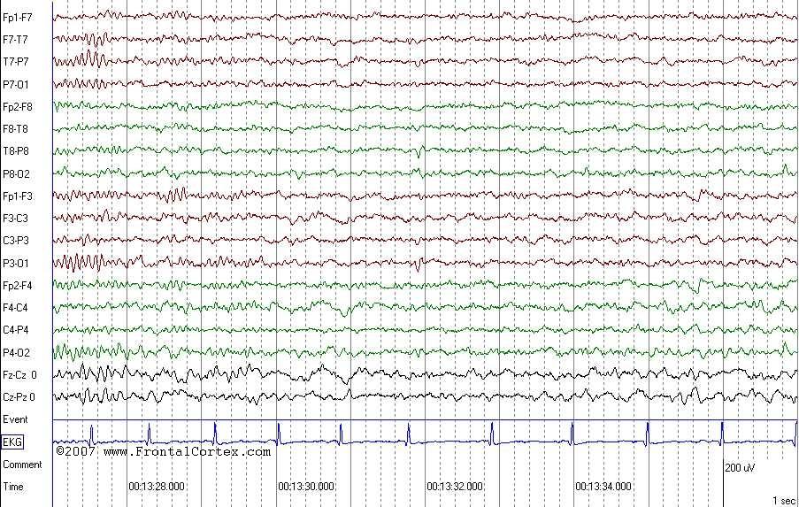
A 9 year-old girl presents to your office accompanied by her parents. The parents state that on 4 occasions over the past several months, the girl has come into their room in the early morning, drooling and having difficulty talking, with a slight facial droop. These symptoms resolved fairly quickly on both occasions. Then, this morning, she had a generalized motor seizure. There is no significant past medical history, and no family history of epilepsy. The patient had a normal birth history, and has consistently met her developmental milestones. Physical exam is normal. An EEG is obtained, and is shown above. Which of the following EEGs is most characteristic of this patient's syndrome?
A)
B)
C)
D)
E)
 | = Go back to the top of the page. |
 | = Reload a different version of this question (). |
 | = Load a random question from the database. |

| = Use this question as a template to create a totally NEW question. |
 | = Enter detailed rating for this question! |
 | = How users like you have rated this question. |
This question was last modified on March 09, 2008.
ANSWERS AND EXPLANATIONS
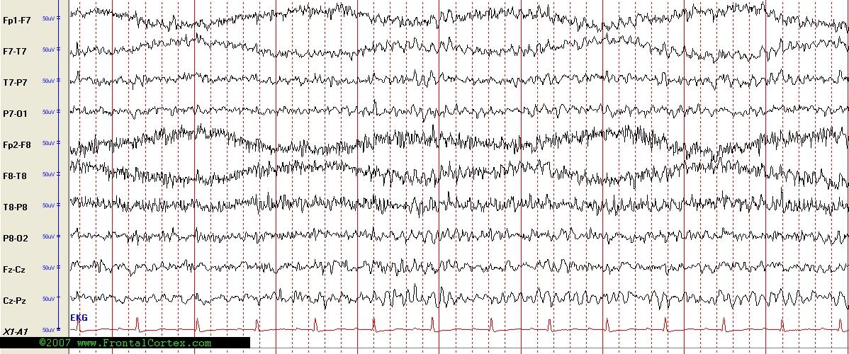
A)
This answer is incorrect.
This child's history is very typical of Benign Childhood Epilepsy with Centrotemporal Spikes (also called BECTS, or "Benign Rolandic Epilepsy"). This syndrome is strongly associated with EEG findings of centrotemporal sharp waves. These sharp waves have a maximal amplitude where the rolandic and sylvian fissures meet. The EEG you selected is a normal EEG, obtained during light drowsiness, with slow roving eye movements. There is no epileptiform activity in this EEG. (See References)
| | |
| | |
| Please log in if you want to rate questions. | |||||
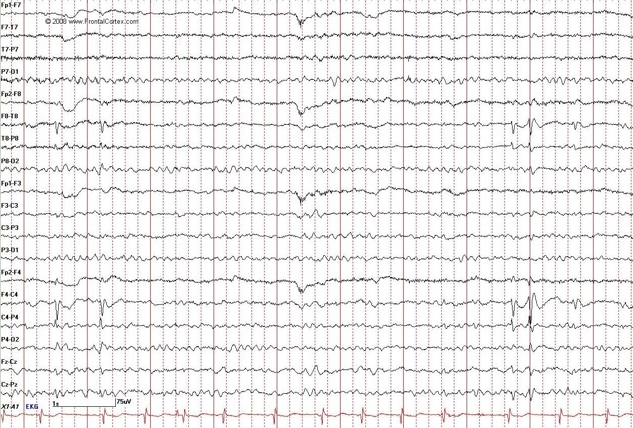
B)
This answer is correct.
This child's history is very typical of Benign Childhood Epilepsy with Centrotemporal Spikes (also called BECTS, or "Benign Rolandic Epilepsy"), the most common focal childhood epilepsy syndrome. Onset of symptoms is between 4 and 12 years, most commonly at age 8 or 9. Seizure types are simple partial motor seizures involving the face, which usually occur during sleep or when the child wakes up. Children are usually brought to clinical attention after a generalized tonic clonic seizure, which represents the secondary generalization of one of these partial motor seizures. There is a family history of epilepsy in 40% of cases. This syndrome is strongly associated with EEG findings of centrotemporal sharp waves. These sharp waves have a maximal amplitude where the rolandic and sylvian fissures meet. Children with this syndrome are otherwise normal neurologically, and typically remit spontaneously. Often, these seizures are not treated. If treated, seizures are usually responsive to monotherapy with most antiepileptic agents. (See References)
| | |
| | |
| Please log in if you want to rate questions. | |||||
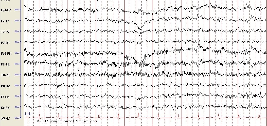
C)
This answer is incorrect.
This child's history is very typical of Benign Childhood Epilepsy with Centrotemporal Spikes (also called BECTS, or "Benign Rolandic Epilepsy"). This syndrome is strongly associated with EEG findings of centrotemporal sharp waves. These sharp waves have a maximal amplitude where the rolandic and sylvian fissures meet. The EEG you selected is a normal EEG with no epileptiform discharges. (See References)
| | |
| | |
| Please log in if you want to rate questions. | |||||
D)
This answer is incorrect.
This child's history is very typical of Benign Childhood Epilepsy with Centrotemporal Spikes (also called BECTS, or "Benign Rolandic Epilepsy"). This syndrome is strongly associated with EEG findings of centrotemporal sharp waves. These sharp waves have a maximal amplitude where the rolandic and sylvian fissures meet. The EEG you selected shows a periodic pattern which would be more consistent with CJD than with BECTS. (See References)
| | |
| | |
| Please log in if you want to rate questions. | |||||
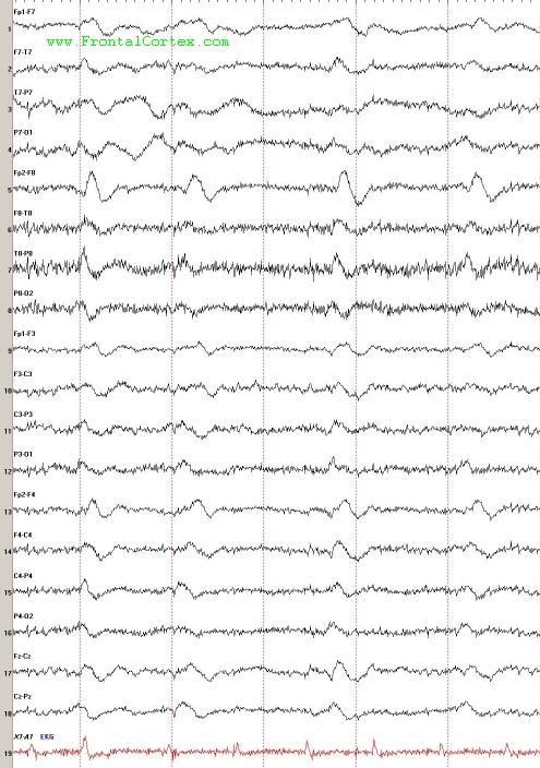
E)
This answer is incorrect.
This child's history is very typical of Benign Childhood Epilepsy with Centrotemporal Spikes (also called BECTS, or "Benign Rolandic Epilepsy"). This syndrome is strongly associated with EEG findings of centrotemporal sharp waves. These sharp waves have a maximal amplitude where the rolandic and sylvian fissures meet. The EEG you selected shows a normal EEG obtained during sleep. There is no epileptiform activity in this EEG. (See References)
| | |
| | |
| Please log in if you want to rate questions. | |||||
References:
| | |
| | |
| Please log in if you want to rate questions. | |||||
FrontalCortex.com -- Neurology Review Questions -- Neurology Boards -- Board Review -- Residency Inservice Training Exam -- RITE Exam Review
pediatric
Pediatric Epilepsy Syndromes 11
Question ID: 030908111
Question written by J. Douglas Miles, (C) 2006-2009, all rights reserved.
Created: 03/09/2008
Modified: 03/09/2008
Estimated Permutations: 8400
User Comments About This Question:
log in to FrontalCortex.com
New to FrontalCortex?
|

![]()
![]()

| | We comply with the HONcode standard for trustworthy health information: verify here. |
Share this page:
 |  |
 |
 |
 |
 |
 |
 |
 |
 |
 |
 |
 |
|
Tuesday, July 01, 2025 at 9:26:06 PM
This site has been visited 49821926 times since June 6 2006
All software and content (C) 2004-2025, FrontalCortex, Inc. unless otherwise specified.
privacy policy Web 2.0 policy disclaimer contact us
All software and content (C) 2004-2025, FrontalCortex, Inc. unless otherwise specified.
privacy policy Web 2.0 policy disclaimer contact us