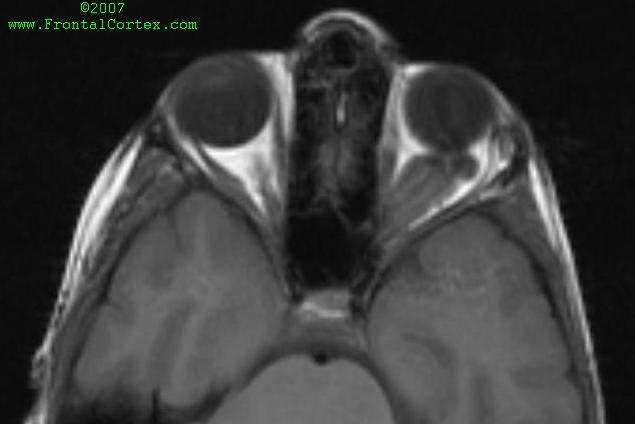
This MRI is most consistent with which of the following diagnoses?
|
| ||||||||||||||||||||||||||||||||||||||||||||||||||||||||||||||||||||||||||||||||||||||||||||||||||||||||||||||||
Neurocutaneous Syndromes 06Topic: ImagingCreated on Saturday, September 22 2007 by jdmiles Last modified on Saturday, September 22 2007.
This MRI is most consistent with which of the following diagnoses? A) Von Hippel-Lindau disease B) Neurofibromatosis type 2 C) Neurofibromatosis type 1 D) Ehlers-Danlos syndrome E) Tuberous sclerosis
This question was last modified on September 22, 2007.
ANSWERS AND EXPLANATIONSA) von Hippel-Lindau disease
| ||||||||||||||||||||||||||||||||||||||||||||||||||||||||||||||||||||||||||||||||||||||||||||||||||||||||||||||||
 |  |  | 
|  |  |
| Please log in if you want to rate questions. | |||||
The MRI shows an optic glioma behind the left eye. Optic gliomas are a common finding in neurofibromatosis type 1 (NF1).
Optic gliomas are not a common finding in neurofibromatosis type 2 (NF2). Acoustic neuromas are more commonly associated with NF2.
(See References) | | |
| | |
| Please log in if you want to rate questions. | |||||
The MRI shows an optic glioma behind the left eye. Optic gliomas are a common finding in neurofibromatosis type 1 (NF1).
Other characteristic features of NF1 include cafe au lait spots, axillary freckling, palpable neurofibromas, Lisch nodules, and bone lesions.
(See References) | | |
| | |
| Please log in if you want to rate questions. | |||||
The MRI shows an optic glioma behind the left eye. Optic gliomas are a common finding in neurofibromatosis type 1 (NF1).
Ehlers-Danlos syndrome (EDS) does not typically present with an optic glioma. Radiographically visible lesions typically associated wtih EDS include large, sometimes multiple aneurysms.
(See References) | | |
| | |
| Please log in if you want to rate questions. | |||||
The MRI shows an optic glioma behind the left eye. Optic gliomas are a common finding in neurofibromatosis type 1 (NF1).
Tuberous sclerosis (TS) does not typically present with an optic glioma. Radiographically visible lesions typically associated wtih TS include subependymal hamartomas and cortical tubers.
(See References) | | |
| | |
| Please log in if you want to rate questions. | |||||
| 1. Rowland, L.P. (Ed) (2000). Merritt's Neurology, 10th Edition. Lippincott Williams & Wilkins, Philadelphia. | |
| 2. Fenichel, G.M. (2005). Clinical Pediatric Neurology, 5th ed. Elsevier, Philadelphia. | |
| 3. Santos, C.C., Miller, V.S., and Roach, E.S. (2004). Neurocutaneous syndromes. In Bradley, W.G., Daroff, R.B., Fenichel, G.M., and Jankovic, J. (Eds.). Neurology in Clinical Practice, Fourth Edition. Butterworth Heinemann, Philadelphia, pp. 1867-1900. | |
| 4. Victor, M., and Ropper, A.H. (2001). Adams and Victor's Principles of Neurology, 7th Edition. McGraw-Hill, New York. | |
| 5. Prayson, R.A., and Goldblum, J.R. (Eds.) (2005). Neuropathology. Elsevier Churchill Livingstone, Philadelphia. |
| | |
| | |
| Please log in if you want to rate questions. | |||||
FrontalCortex.com -- Neurology Review Questions -- Neurology Boards -- Board Review -- Residency Inservice Training Exam -- RITE Exam Review
log in to FrontalCortex.com
New to FrontalCortex?
|

![]()
![]()

| | We comply with the HONcode standard for trustworthy health information: verify here. |
Share this page:
 |  |
 |
 |
 |
 |
 |
 |
 |
 |
 |
 |
 |
|