| Back to "Miscellaneous Demyelinating Diseases" |
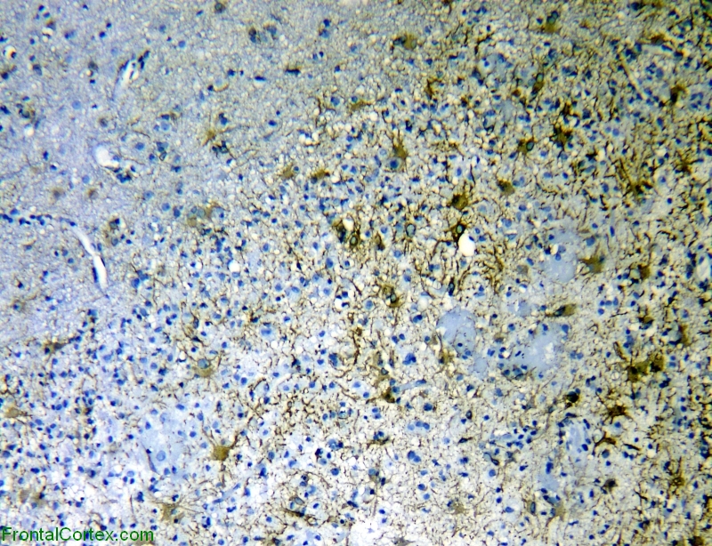
Tumefactive demyelination, immunohistochemical staining for GFAP x 100
Last updated on Monday, April 20 2009 by gliageek
|
| This photograph is taken at the edge of the tumefactive demyelinating lesion. Note the intimate admixture of GFAP-positive reactive astrocytes and GFAP nonreactive macrophages. |
Categories (tags) users associate with this resource 
Please type in an appropriate tag for this item
Click on a tag to find related images, videos, MCQs, and other resources.
Check the boxes next to the tags you consider relevant or enter your own tags in the field below.
You must be logged in to edit tags.
Top 5 tags for this item:
No tags have been created yet for this resource.Please type in an appropriate tag for this item
more tags:
new tag:
log in to FrontalCortex.com
New to FrontalCortex?
|

![]()
![]()

| | We comply with the HONcode standard for trustworthy health information: verify here. |
Share this page:
 |  |
 |
 |
 |
 |
 |
 |
 |
 |
 |
 |
 |
|
Saturday, July 12, 2025 at 1:01:24 AM
This site has been visited 49899671 times since June 6 2006
All software and content (C) 2004-2025, FrontalCortex, Inc. unless otherwise specified.
privacy policy Web 2.0 policy disclaimer contact us
All software and content (C) 2004-2025, FrontalCortex, Inc. unless otherwise specified.
privacy policy Web 2.0 policy disclaimer contact us