| Back to "Miscellaneous Demyelinating Diseases" |
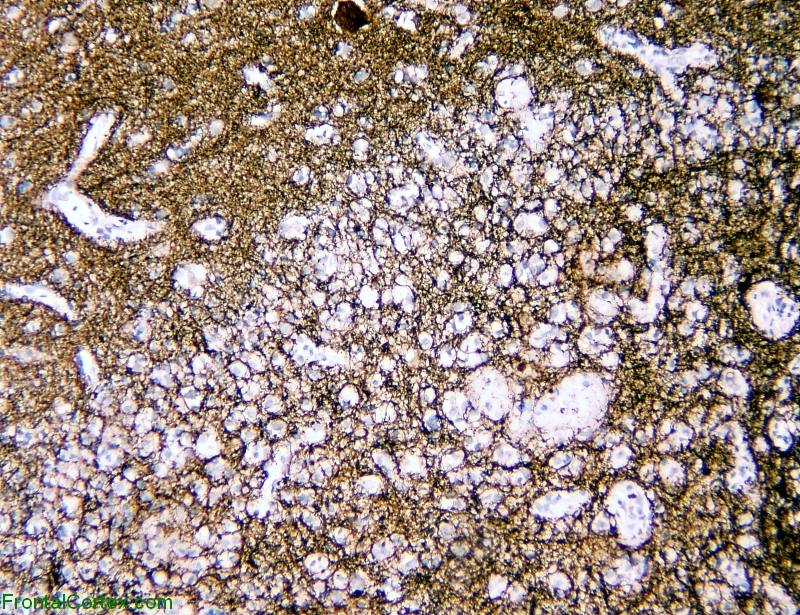
tumefactive demyelination, brain/lesion interface, neurofilament immunostaining x 100
Last updated on Thursday, March 26 2009 by gliageek
|
| Axons are relatively well preserved within tumefactive demyelinating lesions. macrophages stand out as negatively stained well circumscribed discohesive cells within the neurofilament matrix. |
Categories (tags) users associate with this resource 
Please type in an appropriate tag for this item
Click on a tag to find related images, videos, MCQs, and other resources.
Check the boxes next to the tags you consider relevant or enter your own tags in the field below.
You must be logged in to edit tags.
Top 5 tags for this item:
No tags have been created yet for this resource.Please type in an appropriate tag for this item
more tags:
new tag:
log in to FrontalCortex.com
New to FrontalCortex?
|

![]()
![]()

| | We comply with the HONcode standard for trustworthy health information: verify here. |
Share this page:
 |  |
 |
 |
 |
 |
 |
 |
 |
 |
 |
 |
 |
|
Monday, July 14, 2025 at 12:53:31 PM
This site has been visited 49920866 times since June 6 2006
All software and content (C) 2004-2025, FrontalCortex, Inc. unless otherwise specified.
privacy policy Web 2.0 policy disclaimer contact us
All software and content (C) 2004-2025, FrontalCortex, Inc. unless otherwise specified.
privacy policy Web 2.0 policy disclaimer contact us