| Back to "DNT" |
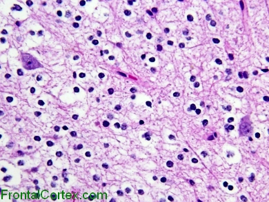
Dysembryoplastic Neuroepithelial Tumor, H&E stain
Last updated on Thursday, April 9 2009 by gliageek
|
| This photograph demonstrates the "specific Glioneuronal element" which characterizes the dysembryoplastic neuroepithelial tumor. Morphologically normal neurons appear to float in a background of oligodendroglia-like cells. Note the absence of perineuronal satellitosis, which is typically seen in oligodendroglial tumors |
Categories (tags) users associate with this resource 
Please type in an appropriate tag for this item
Click on a tag to find related images, videos, MCQs, and other resources.
Check the boxes next to the tags you consider relevant or enter your own tags in the field below.
You must be logged in to edit tags.
Top 5 tags for this item:
No tags have been created yet for this resource.Please type in an appropriate tag for this item
more tags:
new tag:
log in to FrontalCortex.com
New to FrontalCortex?
|

![]()
![]()

| | We comply with the HONcode standard for trustworthy health information: verify here. |
Share this page:
 |  |
 |
 |
 |
 |
 |
 |
 |
 |
 |
 |
 |
|
Saturday, July 12, 2025 at 12:53:31 AM
This site has been visited 49899618 times since June 6 2006
All software and content (C) 2004-2025, FrontalCortex, Inc. unless otherwise specified.
privacy policy Web 2.0 policy disclaimer contact us
All software and content (C) 2004-2025, FrontalCortex, Inc. unless otherwise specified.
privacy policy Web 2.0 policy disclaimer contact us