| Back to "Vascular Disease 1: Reaction to ischemic injury" |
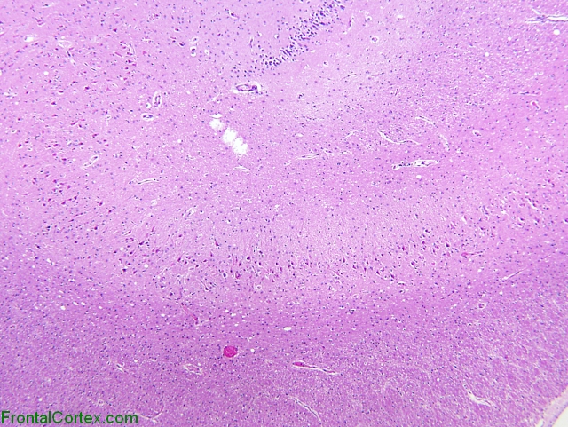
Eosinophilic Neuronal Degeneration, Hippocampus, H&E x40
Last updated on Thursday, April 9 2009 by gliageek
|
| The simple layered structure of the hippocampus compared with the neocortex allows easier interpretation of selective neuronal damage secondary to ischemia. In this photograph, white matter is at the bottom of the slide, a portion of the granular cell layer (dentate gyrus) can be seen at the top of that picture, and hippocampal pyramidal layer occupies the central portion of the photograph running from left to right. |
Categories (tags) users associate with this resource 
Please type in an appropriate tag for this item
Click on a tag to find related images, videos, MCQs, and other resources.
Check the boxes next to the tags you consider relevant or enter your own tags in the field below.
You must be logged in to edit tags.
Top 5 tags for this item:
No tags have been created yet for this resource.Please type in an appropriate tag for this item
more tags:
new tag:
log in to FrontalCortex.com
New to FrontalCortex?
|

![]()
![]()

| | We comply with the HONcode standard for trustworthy health information: verify here. |
Share this page:
 |  |
 |
 |
 |
 |
 |
 |
 |
 |
 |
 |
 |
|
Tuesday, July 15, 2025 at 1:35:55 PM
This site has been visited 49931248 times since June 6 2006
All software and content (C) 2004-2025, FrontalCortex, Inc. unless otherwise specified.
privacy policy Web 2.0 policy disclaimer contact us
All software and content (C) 2004-2025, FrontalCortex, Inc. unless otherwise specified.
privacy policy Web 2.0 policy disclaimer contact us