Basic Neuropathology 9
Topic: Imaging
Created on Monday, February 25 2013 by gliageek
Last modified on Wednesday, February 27 2013.
A) Tabes dorsalis
B) Pellagra
C) Subacute combined degeneration
D) Multiple sclerosis
E) Amyotrophic lateral sclerosis
 | = Go back to the top of the page. |
 | = Reload a different version of this question (). |
 | = Load a random question from the database. |

| = Use this question as a template to create a totally NEW question. |
 | = Enter detailed rating for this question! |
 | = How users like you have rated this question. |
This question was last modified on February 27, 2013.
ANSWERS AND EXPLANATIONS
A) Tabes dorsalis
This answer is incorrect.
As the name implies, tabes dorsalis is characterized by preferential dorsal degeneration of the spinal cord (secondary to Huebner�s arteritis). (See References)
| | |
| | |
| Please log in if you want to rate questions. | |||||
B) Pellagra
This answer is incorrect.
Pellagra, which occurs secondary to riboflavin deficiency, is characterized by chromatolytic changes within anterior horn cells. (See References)
| | |
| | |
| Please log in if you want to rate questions. | |||||
C) Subacute combined degeneration
This answer is incorrect.
Subacute combined degeneration demonstrates preferential damage to the posterior ascending fiber tracts, often with multifocal incomplete corticospinal tract damage. (See References)
| | |
| | |
| Please log in if you want to rate questions. | |||||
D) Multiple sclerosis
This answer is incorrect.
Demyelinating plaques of multiple sclerosis are never confined to anatomically defined tracts (See References)
| | |
| | |
| Please log in if you want to rate questions. | |||||
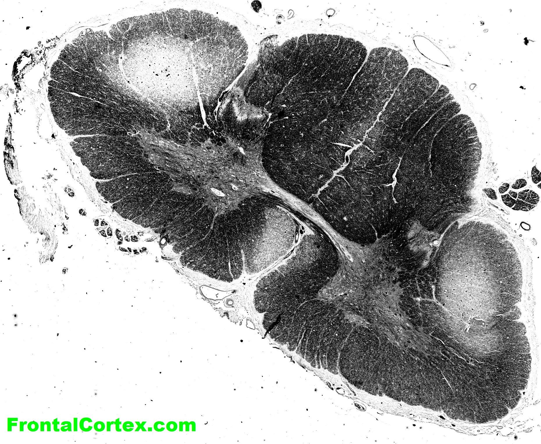
E) Amyotrophic lateral sclerosis
This answer is correct.
Symmetrical degeneration of corticospinal axons occurs secondary to degeneration of cerebrocortical motor neurons in ALS. When axons degenerate, myelin will secondarily be lost. The resulting reactive astrocytosis produces a firm scarlike alteration in the lateral (and anterior) corticospinal tracts or "lateral sclerosis". (See References)
| | |
| | |
| Please log in if you want to rate questions. | |||||
References:
| | |
| | |
| Please log in if you want to rate questions. | |||||
FrontalCortex.com -- Neurology Review Questions -- Neurology Boards -- Board Review -- Residency Inservice Training Exam -- RITE Exam Review
imaging
Basic Neuropathology 9
Question ID: 022513104
Question written by gliageek. (C) FrontalCortex.com 2006-2009, all rights reserved. Created: 02/25/2013
Modified: 02/27/2013
Estimated Permutations: 120
User Comments About This Question:
log in to FrontalCortex.com
New to FrontalCortex?
|

![]()
![]()

| | We comply with the HONcode standard for trustworthy health information: verify here. |
Share this page:
 |  |
 |
 |
 |
 |
 |
 |
 |
 |
 |
 |
 |
|
Friday, June 19, 2026 at 7:05:14 AM
This site has been visited 53447993 times since June 6 2006
All software and content (C) 2004-2026, FrontalCortex, Inc. unless otherwise specified.
privacy policy Web 2.0 policy disclaimer contact us
All software and content (C) 2004-2026, FrontalCortex, Inc. unless otherwise specified.
privacy policy Web 2.0 policy disclaimer contact us