A 534 year-old Renaissance man presents to your office, complaining of numbness.
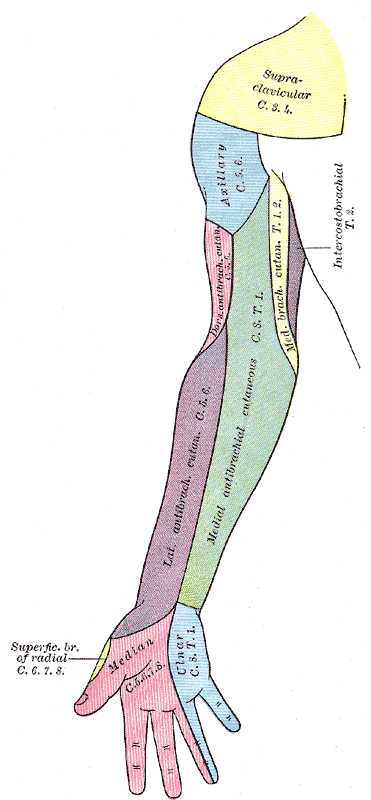
You expertly diagnose him as having a mononeuropathy affecting the medial brachial cutaneous nerve.
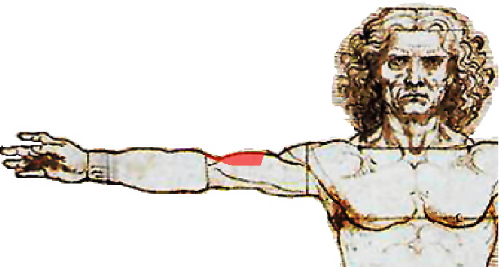
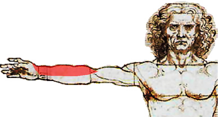
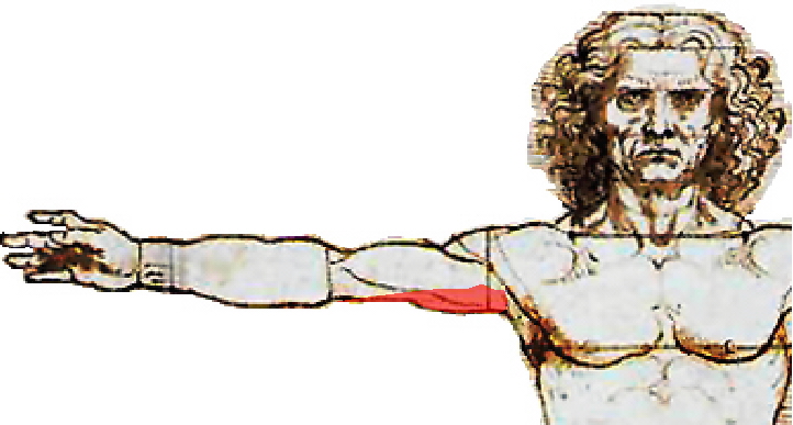
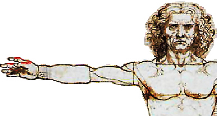
Which image most accurately illustates the pattern of sensory innervation provided by the medial brachial cutaneous nerve?
Sensory innervation of the upper extremity
Topic: Anatomy
Created on Friday, December 22 2006 by jdmiles
Last modified on Sunday, November 16 2008.
A)
B)
C)

D)
E)
This question was last modified on November 16, 2008.
ANSWERS AND EXPLANATIONS
A) 
This answer is incorrect.

The distribution of sensory innervation illustrated in this image best corresponds to the dorsal antibrachial cutaneous nerve. (See References)
 |  |  | 
|  |  |
| Please log in if you want to rate questions. | |||||
B) 
This answer is incorrect.

The distribution of sensory innervation illustrated in this image best corresponds to the lateral antibrachial cutaneous nerve. (See References)
 |  |  | 
|  |  |
| Please log in if you want to rate questions. | |||||
C) 
This answer is correct.

The following image illustrates the distribution of sensory innervation of some of the named nerves in the upper extremity.
 |  |  | 
|  |  |
| Please log in if you want to rate questions. | |||||
D) 
This answer is incorrect.

The distribution of sensory innervation illustrated in this image best corresponds to the intercostobrachial nerve. (See References)
 |  |  | 
|  |  |
| Please log in if you want to rate questions. | |||||
E) 
This answer is incorrect.

The distribution of sensory innervation illustrated in this image best corresponds to the superficial branch of radial nerve. (See References)
 |  |  | 
|  |  |
| Please log in if you want to rate questions. | |||||
References:
| 1. Gray, Henry. (1918). Anatomy of the human body 20th ed. Philadelphia: Lea & Febiger. (ASIN:B000TW11G6) | Advertising: |
 |  |  | 
|  |  |
| Please log in if you want to rate questions. | |||||
FrontalCortex.com -- Neurology Review Questions -- Neurology Boards -- Board Review -- Residency Inservice Training Exam -- RITE Exam Review
anatomy
Sensory innervation of the upper extremity
Question ID: 810200601
Question written by J. Douglas Miles, (C) 2006-2009, all rights reserved.
Created: 12/22/2006
Modified: 11/16/2008
Estimated Permutations: 0


