Toxicity and Characteristic Pathology 02
Topic: Pathology
Created on Wednesday, September 24 2008 by jdmiles
Last modified on Wednesday, September 24 2008.

Courtesy of Dr. Mark Cohen
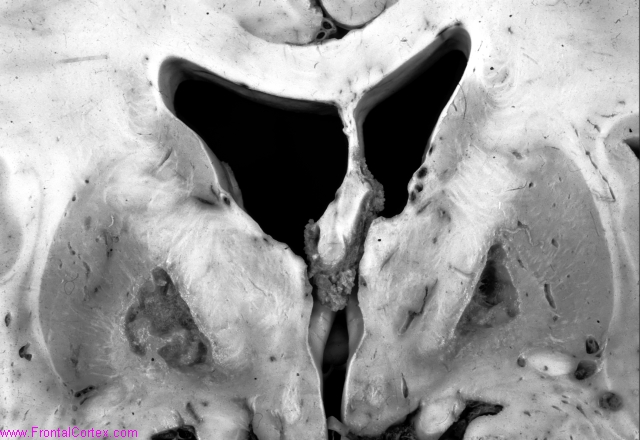
A 44 year old man in previously good health is found at home, comatose and with bright red skin. After a few days in the ICU, the patient expires. An autopsy is performed. The brain is shown in the image above.
What was the cause of death?
This question was created on September 24, 2008 by jdmiles.
This question was last modified on September 24, 2008.
ANSWERS AND EXPLANATIONS
A) Parkinson disease
This answer is incorrect.
The image shows lesions in the globus pallidus bilaterally. This is not the typical lesion seen in Parkinson disease, nor is this young patient's history typical of Parkinson disease. (
See References)
|
 |  |  | 
|  |  |
| Please log in if you want to rate questions. |
B) Carbon monoxide toxicity
This answer is correct.
Bilateral globus pallidus lesions are a common finding in carbon monoxide poisoning. (
See References)
|
 |  |  | 
|  |  |
| Please log in if you want to rate questions. |
C) Ruptured cerebral aneurysm
This answer is incorrect.
The image shows bilateral lesions in the globus pallidus. This is characteristic of carbon monoxide poisoning. Bilateral pallidal aneurysms would be unlikely. (
See References)
|
 |  |  | 
|  |  |
| Please log in if you want to rate questions. |
D) Hallervorden-Spatz disease
This answer is incorrect.
Pantothenate kinase-associated neurodegeneration (PKAN), formerly called Hallervorden-Spatz disease, is a genetic disorder associated with a mutation of the gene coding for pantothenate kinase (PANK2) gene. Patients with PKAN may have iron deposition in the globus pallidus bilaterally. However, what is seen in this patient's brain are necrotic lesions in the pallidi, more characteristic of another disorder.
The name "Hallevorden-Spatz" is no longer used, as both Spatz and Hallevorden were involved in research which involved the euthanasia of victims of the Nazi regime. (
See References)
|
 |  |  | 
|  |  |
| Please log in if you want to rate questions. |
E) Ethylene glycol
This answer is incorrect.
This image shows lesions in the globus pallidus bilaterally. Such lesions have been reported after acute ethylene glycol intoxication. However, the patient's presentation (e.g., red skin) are suggestive of another etiology. (
See References)
|
 |  |  | 
|  |  |
| Please log in if you want to rate questions. |
References:
| 1. Prayson, R.A., and Goldblum, J.R. (Eds.) (2005). Neuropathology. Elsevier Churchill Livingstone, Philadelphia. (ISBN:0443066582)
| Advertising:
|
| 2. Lapresle, J., and Fardeau, M. (1967). "The central nervous system and carbon monoxide poisoning. II. Anatomical study of brain lesions following intoxication with carbon monixide (22 cases)." Prog Brain Res, 24 31-74. (PMID:6075035)
|  |
| 3. Weydt, P. (2000). "Biomedical centre memorial to victims of Nazi research." Nature, 403(6772) 816. (PMID:10733384) |  |
| 4. Caparros-Lefebvre, D., Policard, J., Sengler, C., Benabdallah, E., Colombani, S., and Rigal, M. (2005). "Bipallidal haemorrhage after ethylene glycol intoxication." Neuroradiology, 47(2) 105-7. (PMID:15714272) |  |
|
 |  |  | 
|  |  |
| Please log in if you want to rate questions. |
FrontalCortex.com -- Neurology Review Questions -- Neurology Boards -- Board Review -- Residency Inservice Training Exam -- RITE Exam Review
pathology
Toxicity and Characteristic Pathology 02
Question ID: 092408153
Question written by J. Douglas Miles, (C) 2006-2009, all rights reserved.
Created: 09/24/2008
Modified: 09/24/2008
Estimated Permutations: 600


