| Back to "Tuberous Sclerosis, coronal section demonstrating numerous subependymal nodules and a subependymal giant cell tumor in the septum pellucidum" |
Pediatric Brain Tumor Pathology 01
Topic: Pathology
Created on Sunday, February 24 2008 by jdmiles
Last modified on Sunday, February 24 2008.
A) Tumors of this type often cause hydrocephalus by obstructing the foramen of Magendie
B) This is an oligodendroglioma
C) This is a glioblastoma
D) This patient likely has neurofibromatosis type I
E) This tumor is classified as WHO Grade I
 | = Go back to the top of the page. |
 | = Reload a different version of this question (). |
 | = Load a random question from the database. |

| = Use this question as a template to create a totally NEW question. |
 | = Enter detailed rating for this question! |
 | = How users like you have rated this question. |
This question was last modified on February 24, 2008.
ANSWERS AND EXPLANATIONS
A) Tumors of this type often cause hydrocephalus by obstructing the foramen of Magendie
This answer is incorrect.
This tumor does often cause hydrocephalus. However, tumors of this type typically occur in the lateral ventricle, making them more likely to occlude the foramen of Munro than the foramen of Magendie. (See References)
| | |
| | |
| Please log in if you want to rate questions. | |||||
B) This is an oligodendroglioma
This answer is incorrect.
This tumor does not have an appearance typical of an oligodendroglioma. Oligodendrogliomas classically have a "fried egg" appearance of dense cells. (See References)
| | |
| | |
| Please log in if you want to rate questions. | |||||
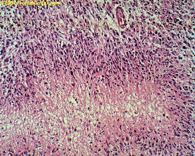
C) This is a glioblastoma
This answer is incorrect.
This tumor does not have an appearance typical of a glioblastoma. Glioblastomas are rapidly-growing tumors. On microscopy, they tend to show pseudopallisading and necrosis. An example is shown below:
| | |
| | |
| Please log in if you want to rate questions. | |||||
D) This patient likely has neurofibromatosis type I
This answer is incorrect.
This tumor is associated with tuberous sclerosis, not neurofibromatosis. (See References)
| | |
| | |
| Please log in if you want to rate questions. | |||||
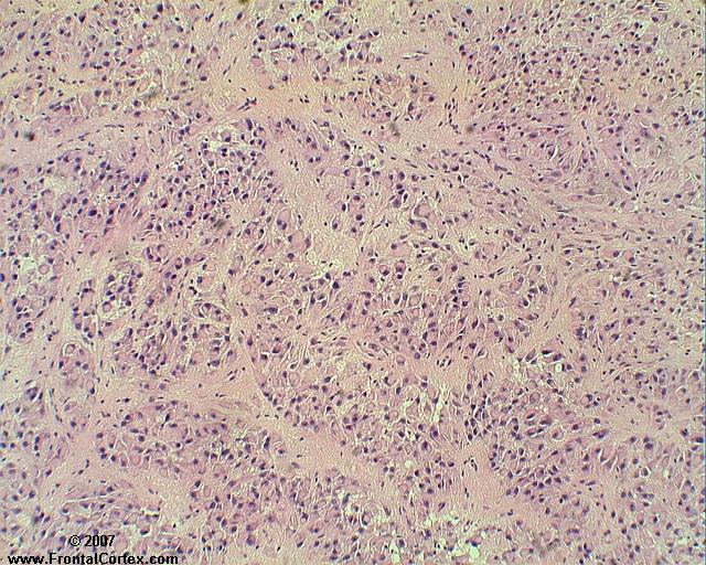
E) This tumor is classified as WHO Grade I
This answer is correct.
This tumor is a subependymal giant cell astrocytoma (SEGA). These tumors are typically long and sausagelike in appearance, often occurring in the lateral ventricle. They may cause hydrocephalus by obstructing the foramen of Munro. They are the most frequent neoplasm in children with tuberous sclerosis complex (TSC), and most SEGAs are associated with TSC. Calcification is typical. This is a slow-growing, WHO Grade I tumor. Pathologic appearance is as shown, with many astrocytes with abundant cytoplasm, often arranged in pseudorosettes. Some tumor cells may appear neuronal, and have prominent nucleoli. (See References)
| | |
| | |
| Please log in if you want to rate questions. | |||||
References:
| | |
| | |
| Please log in if you want to rate questions. | |||||
FrontalCortex.com -- Neurology Review Questions -- Neurology Boards -- Board Review -- Residency Inservice Training Exam -- RITE Exam Review
pathology
Pediatric Brain Tumor Pathology 01
Question ID: 022408120
Question written by J. Douglas Miles, (C) 2006-2009, all rights reserved.
Created: 02/24/2008
Modified: 02/24/2008
Estimated Permutations: 60480
User Comments About This Question:
log in to FrontalCortex.com
New to FrontalCortex?
|

![]()
![]()

| | We comply with the HONcode standard for trustworthy health information: verify here. |
Share this page:
 |  |
 |
 |
 |
 |
 |
 |
 |
 |
 |
 |
 |
|
Friday, July 11, 2025 at 10:34:05 PM
This site has been visited 49898399 times since June 6 2006
All software and content (C) 2004-2025, FrontalCortex, Inc. unless otherwise specified.
privacy policy Web 2.0 policy disclaimer contact us
All software and content (C) 2004-2025, FrontalCortex, Inc. unless otherwise specified.
privacy policy Web 2.0 policy disclaimer contact us