| Back to "Viral Infections" |
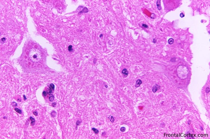
Rabies - Negri Bodies H&E
Last updated on Thursday, February 5 2009 by jdmiles
|
| This micrograph depicts the histopathologic changes associated with rabies encephalitis prepared using an H&E stain. Note the Negri bodies, which are cellular inclusions found most frequently in the pyramidal cells of Ammon's horn, and the Purkinje cells of the cerebellum. They are also found in the cells of the medulla and various other ganglia. Courtesy of CDC / Dr. Daniel P. Perl This image is in the public domain and thus free of any copyright restrictions. As a matter of courtesy we request that the content provider be credited and notified in any public or private usage of this image. |
Related images:
|
| Rabies - Negri Bodies H&E with arrow The unnecessarily large green arrow points to one of several Negri bodies visible in this image. They are dark pink blobs in the cytoplasm. |
Categories (tags) users associate with this resource 
Please type in an appropriate tag for this item
Click on a tag to find related images, videos, MCQs, and other resources.
Check the boxes next to the tags you consider relevant or enter your own tags in the field below.
You must be logged in to edit tags.
Top 5 tags for this item:
No tags have been created yet for this resource.Please type in an appropriate tag for this item
more tags:
new tag:
log in to FrontalCortex.com
New to FrontalCortex?
|

![]()
![]()

| | We comply with the HONcode standard for trustworthy health information: verify here. |
Share this page:
 |  |
 |
 |
 |
 |
 |
 |
 |
 |
 |
 |
 |
|
Friday, July 11, 2025 at 10:21:50 PM
This site has been visited 49898293 times since June 6 2006
All software and content (C) 2004-2025, FrontalCortex, Inc. unless otherwise specified.
privacy policy Web 2.0 policy disclaimer contact us
All software and content (C) 2004-2025, FrontalCortex, Inc. unless otherwise specified.
privacy policy Web 2.0 policy disclaimer contact us