| Back to "Vascular Disease 4: Other topics in vascular disease" |
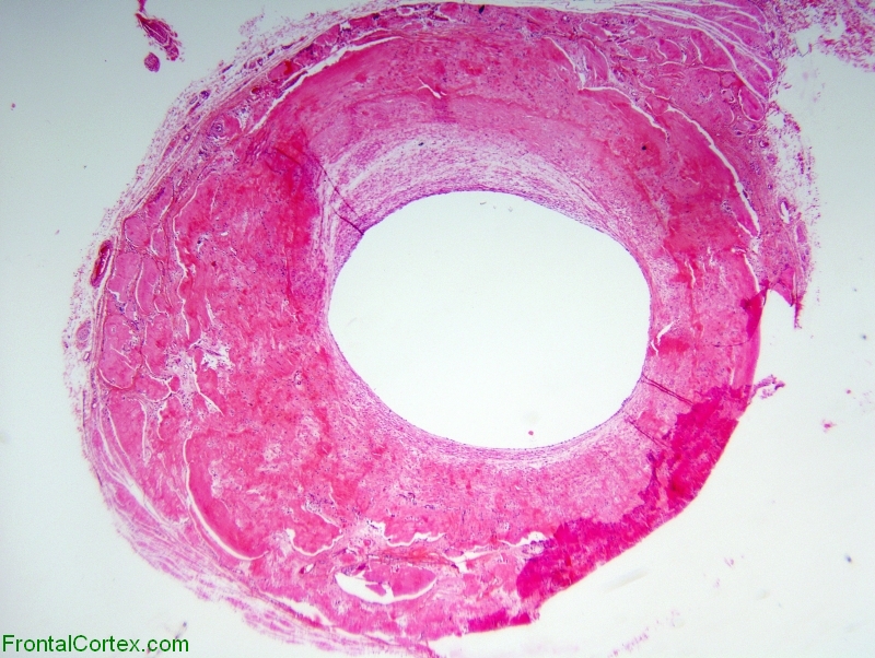
Amyloid angiopathy involving temporal artery, H&E stain x 20
Last updated on Thursday, April 9 2009 by gliageek
|
| Hyalin thickening of the arterial wall with loss of normal cellular components. Unlike leptomeningeal blood vessels where amyloid angiopathy is nearly always due to vascular involvement by beta amyloid, temporal arteries demonstrate vascular amyloid secondary to proteins associated with systemic amyloidoses. This patient was subsequently found to have multiple myeloma with systemic light chain amyloidosis. |
Categories (tags) users associate with this resource 
Please type in an appropriate tag for this item
Click on a tag to find related images, videos, MCQs, and other resources.
Check the boxes next to the tags you consider relevant or enter your own tags in the field below.
You must be logged in to edit tags.
Top 5 tags for this item:
No tags have been created yet for this resource.Please type in an appropriate tag for this item
more tags:
new tag:
log in to FrontalCortex.com
New to FrontalCortex?
|

![]()
![]()

| | We comply with the HONcode standard for trustworthy health information: verify here. |
Share this page:
 |  |
 |
 |
 |
 |
 |
 |
 |
 |
 |
 |
 |
|
Friday, July 18, 2025 at 5:08:06 PM
This site has been visited 49961314 times since June 6 2006
All software and content (C) 2004-2025, FrontalCortex, Inc. unless otherwise specified.
privacy policy Web 2.0 policy disclaimer contact us
All software and content (C) 2004-2025, FrontalCortex, Inc. unless otherwise specified.
privacy policy Web 2.0 policy disclaimer contact us