| Back to "Inactive demyelinated plaque, H&E stain section" |
Inactive demyelinated plaque, H&E stain section
Last updated on Friday, March 27 2009 by jdmiles
|
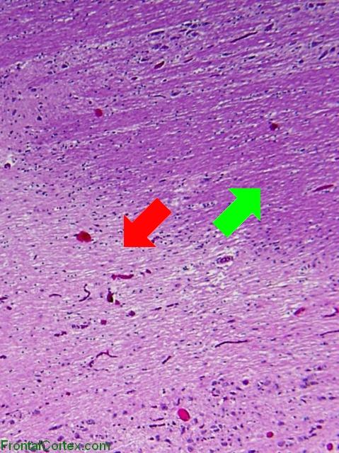
| This is an H&E preparation of an older, inactive demyelinated plaque from the brain of a person with MS. There is no active demyelination going on in this plaque. In this image, we see the border between the plaque (pale, indicated by the unnecessarily large red arrow) and normal neuropil (darker, indicated by the unnecessarily large green arrow). Note that, while the plaque is pale, indicating a lack of myelin, the overall structure appears very similar to that of the normal neuropil, as axons remain intact. |
Categories (tags) users associate with this resource 
Please type in an appropriate tag for this item
Click on a tag to find related images, videos, MCQs, and other resources.
Check the boxes next to the tags you consider relevant or enter your own tags in the field below.
You must be logged in to edit tags.
Top 5 tags for this item:
No tags have been created yet for this resource.Please type in an appropriate tag for this item
more tags:
new tag:
log in to FrontalCortex.com
New to FrontalCortex?
|

![]()
![]()

| | We comply with the HONcode standard for trustworthy health information: verify here. |
Share this page:
 |  |
 |
 |
 |
 |
 |
 |
 |
 |
 |
 |
 |
|
Sunday, May 05, 2024 at 1:00:00 PM
This site has been visited 45930317 times since June 6 2006
All software and content (C) 2004-2024, FrontalCortex, Inc. unless otherwise specified.
privacy policy Web 2.0 policy disclaimer contact us
All software and content (C) 2004-2024, FrontalCortex, Inc. unless otherwise specified.
privacy policy Web 2.0 policy disclaimer contact us