| Back to "Neuromuscular disorders 1: Myopathies and Dystrophies" |
Example of trichrome stain
Last updated on Friday, February 6 2009 by jdmiles
|
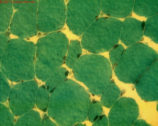
| This is an example of a muscle specimen stained with Gomori trichrome. On trichrome, muscle typically shows up as a greenish blue (or in this case, just green). This particular muscle biopsy is an example of nemaline rod myopathy. The nemaline rods are tiny dark spots visible in most of the muscle fibers. This is an example of something we'd see on trichrome that we wouldn't see on H&E. |
Related images:
|
| Gomori Trichrome Nemaline Rods 2 A Gomori trichrome stain of skeletal muscle from an individual with nemaline rod myopathy. The rods themselves show up as small dark dots on trichrome (highlighted here by the red circles). Note that these rods typically do not show up on H&E staining. |
Categories (tags) users associate with this resource 
Please type in an appropriate tag for this item
Click on a tag to find related images, videos, MCQs, and other resources.
Check the boxes next to the tags you consider relevant or enter your own tags in the field below.
You must be logged in to edit tags.
Top 5 tags for this item:
No tags have been created yet for this resource.Please type in an appropriate tag for this item
more tags:
new tag:
log in to FrontalCortex.com
New to FrontalCortex?
|

![]()
![]()

| | We comply with the HONcode standard for trustworthy health information: verify here. |
Share this page:
 |  |
 |
 |
 |
 |
 |
 |
 |
 |
 |
 |
 |
|
Tuesday, July 01, 2025 at 8:21:51 PM
This site has been visited 49821594 times since June 6 2006
All software and content (C) 2004-2025, FrontalCortex, Inc. unless otherwise specified.
privacy policy Web 2.0 policy disclaimer contact us
All software and content (C) 2004-2025, FrontalCortex, Inc. unless otherwise specified.
privacy policy Web 2.0 policy disclaimer contact us