|
| There are 484 questions on various topics in Neurology in the FrontalCortex neurology question bank. |
|
Cerebrovascular Anatomy 02
Topic: AnatomyCreated on Friday, October 12 2007 by jdmiles
Last modified on Friday, October 12 2007.
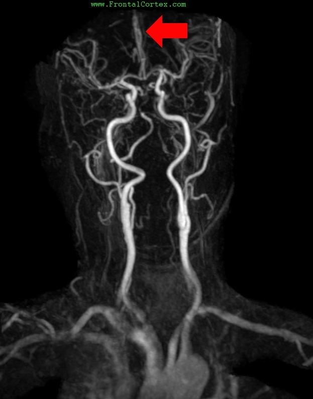
An occlusion of the indicated vessel would result in which of the following deficits?
This question was created on October 12, 2007 by jdmiles.
This question was last modified on October 12, 2007.
ANSWERS AND EXPLANATIONS
A) Anton syndrome
This answer is incorrect.
The arrow in this gadolinium bolus MR angiogram indicates the left anterior cerebral artery. An occlusion of this vessel would produce hemiparesis of the lower extremity, sparing the face and upper extremity. Anton syndrome (denial of blindness by a person who obviously cannot see) is associated with lesions in the visual association cortex. ( See References)
|
 |  |  | 
|  |  | | Please log in if you want to rate questions. |
B) Hemiparesis which affects the lower extremity more than the upper extremity
This answer is correct.
The arrow in this gadolinium bolus MR angiogram indicates the left anterior cerebral artery. An occlusion of this vessel would produce hemiparesis of the lower extremity, sparing the face and upper extremity. ( See References)
|
 |  |  | 
|  |  | | Please log in if you want to rate questions. |
C) Hemiparesis which affects the upper extremity more than the lower extremity
This answer is incorrect.
The arrow in this gadolinium bolus MR angiogram indicates the left anterior cerebral artery. An occlusion of this vessel would produce hemiparesis of the lower extremity, sparing the face and upper extremity. ( See References)
|
 |  |  | 
|  |  | | Please log in if you want to rate questions. |
D) A facial droop
This answer is incorrect.
The arrow in this gadolinium bolus MR angiogram indicates the left anterior cerebral artery. An occlusion of this vessel would produce hemiparesis of the lower extremity, sparing the face and upper extremity. ( See References)
|
 |  |  | 
|  |  | | Please log in if you want to rate questions. |
E) Global aphasia
This answer is incorrect.
The arrow in this gadolinium bolus MR angiogram indicates the left anterior cerebral artery. An occlusion of this vessel would produce hemiparesis of the lower extremity, sparing the face and upper extremity. ( See References)
|
 |  |  | 
|  |  | | Please log in if you want to rate questions. |
References:
| 1. Sandhu, J.S., and Wakhloo, A.K. (2004). Neuroangiographic anatomy and common cerebrovascular diseases. In Bradley, W.G., Daroff, R.B., Fenichel, G.M., and Jankovic, J. (Eds.). Neurology in Clinical Practice, Fourth Edition. Butterworth Heinemann, Philadelphia, pp. 625-643 (ISBN:0750674695). | Advertising:
|
| 2. David L. Felten, Ralph F., Md. Jozefowicz, Frank H., Md. Netter, . . ICON Learning Systems (ISBN:1929007167) | Advertising:
|
| 3. Duane E. Haines; special contributions by John A. Lancon; illustrator, M. P. Schenk; photographer, G. W. Armstrong. Neuroanatomy: an atlas of structures, sections, and systems. Philadelphia : Lippincott Williams & Wilkins, c2004. (ISBN:0781746779) | Advertising:
|
|
 |  |  | 
|  |  | | Please log in if you want to rate questions. |
FrontalCortex.com -- Neurology Review Questions -- Neurology Boards -- Board Review -- Residency Inservice Training Exam -- RITE Exam Review
anatomy
Cerebrovascular Anatomy 02
Question ID: 101207048
Question written by J. Douglas Miles, (C) 2006-2009, all rights reserved.
Created: 10/12/2007
Modified: 10/12/2007
Estimated Permutations: 1800
|
|
|
|


