An occlusion of the indicated vessel would result in which of the following deficits?
|
| |||||||||||||||||||||||||||||||||||||||||||||||||||||||||||||||||||||||||||||||||||||||||||||||||||||||||||||||
Cerebrovascular Anatomy 02Topic: AnatomyCreated on Friday, October 12 2007 by jdmiles Last modified on Friday, October 12 2007.
An occlusion of the indicated vessel would result in which of the following deficits? A) A facial droop B) Balint syndrome C) Hemineglect D) Hemiparesis which affects the lower extremity more than the upper extremity E) Global aphasia
This question was last modified on October 12, 2007.
ANSWERS AND EXPLANATIONSA) A facial droop
| |||||||||||||||||||||||||||||||||||||||||||||||||||||||||||||||||||||||||||||||||||||||||||||||||||||||||||||||
 |  |  | 
|  |  |
| Please log in if you want to rate questions. | |||||
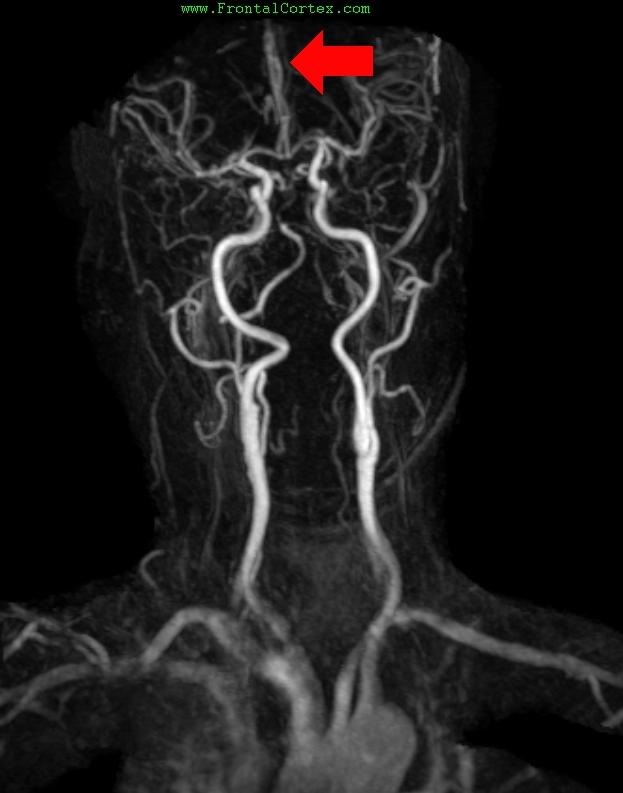
The arrow in this gadolinium bolus MR angiogram indicates the left anterior cerebral artery. An occlusion of this vessel would produce hemiparesis of the lower extremity, sparing the face and upper extremity.
Balint syndrome (a constellation including psychic paralysis of gaze, optic ataxia, and visual inattention) is associated with bilateral lesions in the parietal-occipital vascular border zones.
(See References) | | |
| | |
| Please log in if you want to rate questions. | |||||
The arrow in this gadolinium bolus MR angiogram indicates the left anterior cerebral artery. An occlusion of this vessel would produce hemiparesis of the lower extremity, sparing the face and upper extremity.
Hemineglect is seen in people with lesions of the left parietal cortex.
(See References) | | |
| | |
| Please log in if you want to rate questions. | |||||
| | |
| | |
| Please log in if you want to rate questions. | |||||
| | |
| | |
| Please log in if you want to rate questions. | |||||
| 1. Sandhu, J.S., and Wakhloo, A.K. (2004). Neuroangiographic anatomy and common cerebrovascular diseases. In Bradley, W.G., Daroff, R.B., Fenichel, G.M., and Jankovic, J. (Eds.). Neurology in Clinical Practice, Fourth Edition. Butterworth Heinemann, Philadelphia, pp. 625-643 (ISBN:0750674695). | Advertising: |
| 2. David L. Felten, Ralph F., Md. Jozefowicz, Frank H., Md. Netter, . . ICON Learning Systems (ISBN:1929007167) | Advertising: |
| 3. Duane E. Haines; special contributions by John A. Lancon; illustrator, M. P. Schenk; photographer, G. W. Armstrong. Neuroanatomy: an atlas of structures, sections, and systems. Philadelphia : Lippincott Williams & Wilkins, c2004. (ISBN:0781746779) | Advertising: |
| | |
| | |
| Please log in if you want to rate questions. | |||||
FrontalCortex.com -- Neurology Review Questions -- Neurology Boards -- Board Review -- Residency Inservice Training Exam -- RITE Exam Review
log in to FrontalCortex.com
New to FrontalCortex?
|

![]()
![]()

| | We comply with the HONcode standard for trustworthy health information: verify here. |
Share this page:
 |  |
 |
 |
 |
 |
 |
 |
 |
 |
 |
 |
 |
|