Leukodystrophies 01
Topic: ImagingCreated on Saturday, December 8 2007 by jdmiles
Last modified on Saturday, December 8 2007.
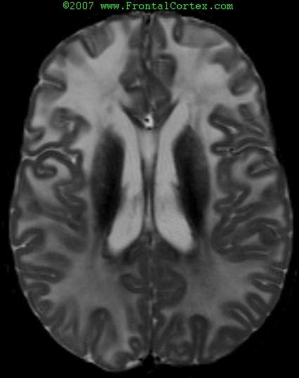
During the workup of a child with a developmental delay, you obtain the above MRI. This image is most consistent with which of the following diagnoses?
This question was created on December 08, 2007 by jdmiles.
This question was last modified on December 08, 2007.
ANSWERS AND EXPLANATIONS
A) Alexander disease
This answer is correct.
The findings in this MRI are typical for Alexander disease. MRI criteria for the diagnosis of Alexander disease include: - Extensive, frontally-predominant cerebral white matter changes
- A periventricular rim which is bright on T1 and dark on T2
- Brainstem abnormalities
- Abnormalities of hte basal ganglia and thalami
- Contrast enhancement of particular gray and white matter structures.
Four of 5 criteria should be met to make the diagnosis based on imaging.
( See References)
|
 |  |  | 
|  |  | | Please log in if you want to rate questions. |
B) Krabbe disease
This answer is incorrect.
In Krabbe disease, the initial lesions to appear hyperintense on MRI tend to be in the basal ganglia and periventricular white matter, and appear symmetric. Subcortical U fibers are spared until late in the disease. The frontal white matter does not tend to be preferentially affected, as it is in the image shown.
( See References)
|
| | |
| | | | Please log in if you want to rate questions. |
C) Zellweger syndrome
This answer is incorrect.
Typical findings in Zellweger syndrome include abnormal gyration and diffuse demyelination. This image shows a frontally-predominant demyelination, which is not characteristic of Zellweger syndrome.
( See References)
|
| | |
| | | | Please log in if you want to rate questions. |
D) Normal variant
This answer is incorrect.
This MRI shows hyperintense lesions in the frontal white matter. This is not normal.
( See References)
|
| | |
| | | | Please log in if you want to rate questions. |
E) Adrenoleukodystrophy
This answer is incorrect.
Adrenoleukodystrophy typically has a characteristic pattern on MRI, in which the occipital and parietal white matter is affected first. As the disease progresses, the leukodystrophy moves anteriorly and caudally. There are atypical cases in which the anterior white matter is affected first; however, classically, the initial demyelination occurs in peritrigonal areas.
( See References)
|
| | |
| | | | Please log in if you want to rate questions. |
References:
| 1. Fenichel, G.M. (2005). Clinical Pediatric Neurology, 5th ed. Elsevier, Philadelphia.
| |
| 2. Robert I. Grossman, David M. Yousem. Neuroradiology: the requisites. Philadelphia, Pa. : Mosby, c2003. (ISBN:978032300508X)
| Advertising:
|
| 3. Sklar, E.M.L., Ruiz, A., Quencer, R.M., and Falcone, S.F. (2004). Structural neuroimaging. In Bradley, W.G., Daroff, R.B., Fenichel, G.M., and Jankovic, J. (Eds.). Neurology in Clinical Practice, Fourth Edition. Butterworth Heinemann, Philadelphia, pp. 521-597 (ISBN:0750674695).
| Advertising:
|
| 4. Cheon, J., Kim, I., Hwang, Y.S., Kim, K.J., Wang, K., Cho, B., Chi, J.G., Kim, C.J., Kim, W.S., and Yeon, K.M. (2002). "Leukodystrophy in children: a pictorial review of MR imaging features." Radiographics, 22(3) 461-76. (PMID:12006681) |  |
| 5. Magnaldi, S. (1991). "[Leukodystrophies: clinical aspects and findings with computerized tomography and magnetic resonance imaging]" Radiol Med (Torino), 82(1-2) 13-26. (PMID:1896564) | |
| 6. van der Knaap, M.S., Naidu, S., Breiter, S.N., Blaser, S., Stroink, H., Springer, S., Begeer, J.C., van Coster, R., Barth, P.G., Thomas, N.H., Valk, J., and Powers, J.M. (2001). "Alexander disease: diagnosis with MR imaging." AJNR Am J Neuroradiol, 22(3) 541-52. (PMID:11237983) | |
|
| | |
| | | | Please log in if you want to rate questions. |
FrontalCortex.com -- Neurology Review Questions -- Neurology Boards -- Board Review -- Residency Inservice Training Exam -- RITE Exam Review
imaging
Leukodystrophies 01
Question ID: 101207183
Question written by J. Douglas Miles, (C) 2006-2009, all rights reserved.
Created: 12/08/2007
Modified: 12/08/2007
Estimated Permutations: 8400
0 user entries
|
|