Adult Brain Tumor Pathology 01
Topic: Pathology
Created on Wednesday, February 4 2009 by jdmiles
Last modified on Wednesday, February 4 2009.
Courtesy of Dr. Mark Cohen
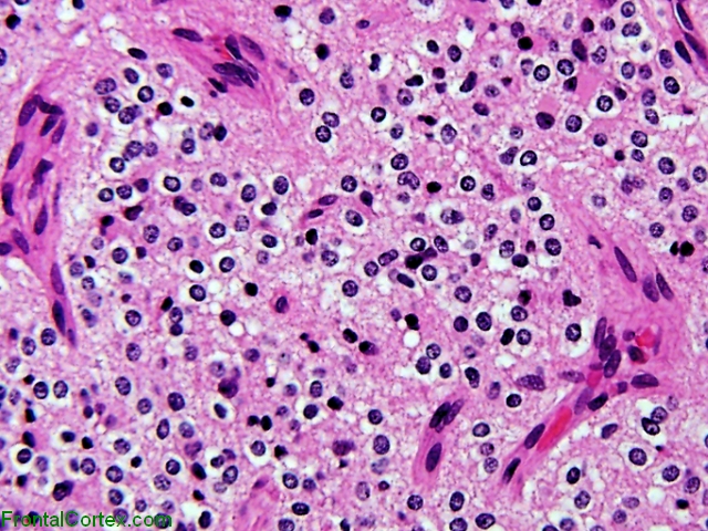
A 54 year old woman presents with slowly worsening seizures over 17 months. Imaging of the head reveals a mass lesion. The lesion is surgically resected, and an H&E preparation of the tumor is shown in the image above.
What kind of tumor is this?
This question was created on February 04, 2009 by jdmiles.
This question was last modified on February 04, 2009.
ANSWERS AND EXPLANATIONS
A) glioblastoma multiforme
This answer is incorrect.
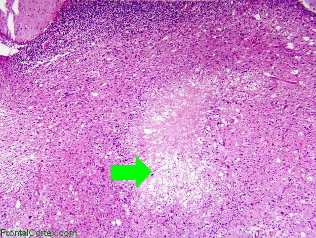
The whorls of cells seen in the pathology from our patient (in the image at the top of this page) are more characteristic of a meningioma than a glioblastoma. Glioblastomas tend to appear more disorganized. They are rapidly-growing tumors which often have areas of necrosis, as shown in the image below (the unnecessarily large green arrow points to the necrotic region):
(
See References)
|
 |  |  | 
|  |  |
| Please log in if you want to rate questions. |
B) pilocytic astrocytoma
This answer is incorrect.
The whorls of cells seen in the pathology from our patient (in the image at the top of this page) are more characteristic of a meningioma than a pilocytic astrocytoma.
Pilocytic astrocytomas are the most common glioma in children, but account for 2% of all primary CNS tumors in all age groups. Pilocytic astrocytomas look more characteristically like the image below:
Courtesy of Dr. Mark Cohen
(
See References)
|
| | |
| | |
| Please log in if you want to rate questions. |
C) medulloblastoma
This answer is incorrect.
The whorls of cells seen in the pathology from our patient (in the image at the top of this page) are more characteristic of a meningioma than a medulloblastoma.
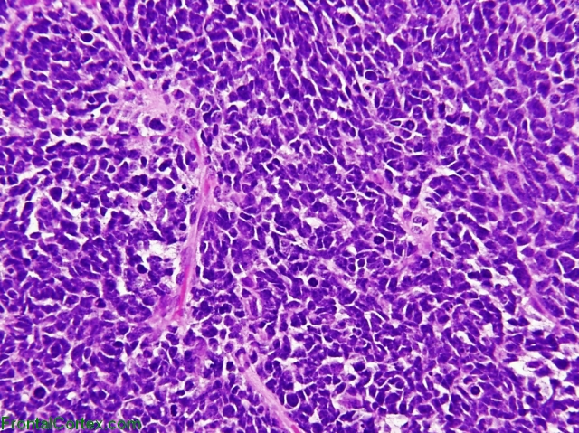
The vast majority of medulloblastomas occur in children. The appearance of medulloblastomas is one of small, blue cells, as shown below:
Courtesy of Dr. Mark Cohen
(
See References)
|
| | |
| | |
| Please log in if you want to rate questions. |
D) oligodendroglioma
This answer is incorrect.
The whorls of cells seen in the pathology from our patient (in the image at the top of this page) are more characteristic of a meningioma than a an oligodendroglioma.
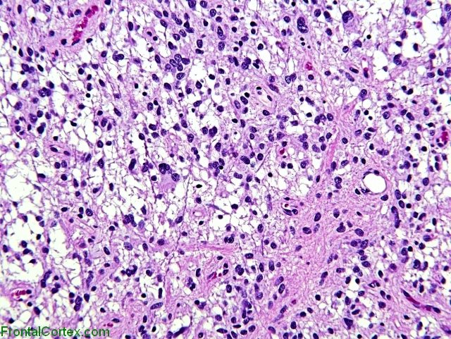
Oligodendrogliomas have a characteristic appearance, with cells that look like a bunch of "fried eggs." An oligodendroglioma is shown in the image below:
Courtesy of Dr. Mark Cohen
(
See References)
|
| | |
| | |
| Please log in if you want to rate questions. |
E) meningioma
This answer is correct.
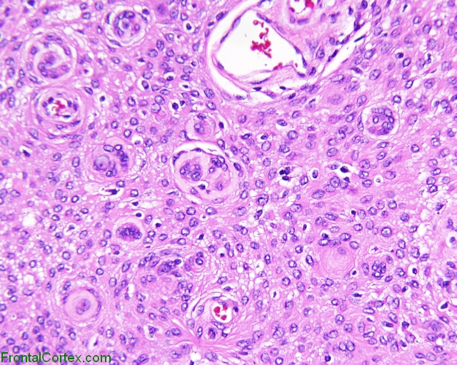
Meningiomas are relatively common non-glial tumors. They are characterized by whorls of meningioepithelial cells, and these whorls are evident in the image shown. Most are curable by total resection.
(
See References)
|
| | |
| | |
| Please log in if you want to rate questions. |
References:
| 1. Prayson, R.A., and Goldblum, J.R. (Eds.) (2005). Neuropathology. Elsevier Churchill Livingstone, Philadelphia. (ISBN:0443066582)
| Advertising:
|
|
| | |
| | |
| Please log in if you want to rate questions. |
FrontalCortex.com -- Neurology Review Questions -- Neurology Boards -- Board Review -- Residency Inservice Training Exam -- RITE Exam Review
pathology
Adult Brain Tumor Pathology 01
Question ID: 020409046
Question written by J. Douglas Miles, (C) 2006-2009, all rights reserved.
Created: 02/04/2009
Modified: 02/04/2009
Estimated Permutations: 120