Vascular Malformations 02
Topic: Pathology
Created on Saturday, April 28 2007 by jdmiles
Last modified on Saturday, April 28 2007.
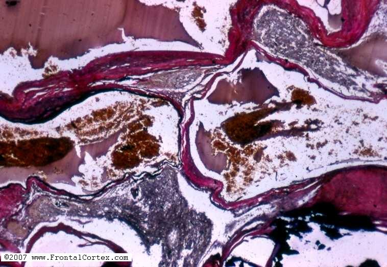
Which of the following statements about the type of lesion seen in this photomicrograph is most accurate?
This question was created on April 28, 2007 by jdmiles.
This question was last modified on April 28, 2007.
ANSWERS AND EXPLANATIONS
A) lesions of this type are dilated veins of the superficial or subcortical vasculature
This answer is incorrect.
The photomicrograph shows an arteriovenous malformation. AVMs are masses of thick-walled blood vessels of varying caliber. They are associated with a 2/3 risk of clinically significant hemorrhage, are associated with a 2%-4% annual risk of acute hemorrhage, account for 1.5%-4% of all brain masses, and have an initial presentation of cerebral hemorrhage in 50% of cases. They are not dilated veins. Venous angiomas are vascular malformations which are dilated veins of the superficial or subcortical vasculature. (
See References)
|
 |  |  | 
|  |  |
| Please log in if you want to rate questions. |
B) this type of lesion consists of dilated capillaries separated by normal brain tissue
This answer is incorrect.
The photomicrograph shows an arteriovenous malformation. AVMs are masses of thick-walled blood vessels of varying caliber. They are associated with a 2/3 risk of clinically significant hemorrhage, are associated with a 2%-4% annual risk of acute hemorrhage, account for 1.5%-4% of all brain masses, and have an initial presentation of cerebral hemorrhage in 50% of cases. Capillary telangiectases consist of dilated capillaries separated by normal brain tissue. (
See References)
|
| | |
| | |
| Please log in if you want to rate questions. |
C) lesions of this type rarely become symptomatic
This answer is incorrect.
The photomicrograph shows an arteriovenous malformation. AVMs are masses of thick-walled blood vessels of varying caliber. They are associated with a 2/3 risk of clinically significant hemorrhage, are associated with a 2%-4% annual risk of acute hemorrhage, account for 1.5%-4% of all brain masses, and have an initial presentation of cerebral hemorrhage in 50% of cases. Capillary telangiectases and venous angiomas are vascular malformations that rarely become symptomatic. (
See References)
|
| | |
| | |
| Please log in if you want to rate questions. |
D) this is a cavernous angioma
This answer is incorrect.
The photomicrograph shows an arteriovenous malformation. AVMs are masses of thick-walled blood vessels of varying caliber. They are associated with a 2/3 risk of clinically significant hemorrhage, are associated with a 2%-4% annual risk of acute hemorrhage, account for 1.5%-4% of all brain masses, and have an initial presentation of cerebral hemorrhage in 50% of cases. (
See References)
|
| | |
| | |
| Please log in if you want to rate questions. |
E) this is an arteriovenous malformation
This answer is correct.
The photomicrograph shows an arteriovenous malformation. AVMs are masses of thick-walled blood vessels of varying caliber. They are associated with a 2/3 risk of clinically significant hemorrhage, are associated with a 2%-4% annual risk of acute hemorrhage, account for 1.5%-4% of all brain masses, and have an initial presentation of cerebral hemorrhage in 50% of cases. (
See References)
|
| | |
| | |
| Please log in if you want to rate questions. |
References:
| 1. Prayson, R.A., and Goldblum, J.R. (Eds.) (2005). Neuropathology. Elsevier, Philadelphia. | |
|
| | |
| | |
| Please log in if you want to rate questions. |
FrontalCortex.com -- Neurology Review Questions -- Neurology Boards -- Board Review -- Residency Inservice Training Exam -- RITE Exam Review
pathology
Vascular Malformations 02
Question ID: 042807149
Question written by J. Douglas Miles, (C) 2006-2009, all rights reserved.
Created: 04/28/2007
Modified: 04/28/2007
Estimated Permutations: 151200