Cerebellar Microanatomy 01
Topic: Pathology
Created on Saturday, November 24 2007 by jdmiles
Last modified on Saturday, November 24 2007.
A) Adult cerebellum
B) Adolescent cerebellum
C) This could be cerebellum from an adult or from an adolescent
D) This is choroid plexus
E) Neonatal cerebellum
 | = Go back to the top of the page. |
 | = Reload a different version of this question (). |
 | = Load a random question from the database. |

| = Use this question as a template to create a totally NEW question. |
 | = Enter detailed rating for this question! |
 | = How users like you have rated this question. |
This question was last modified on November 24, 2007.
ANSWERS AND EXPLANATIONS
A) Adult cerebellum
This answer is incorrect.
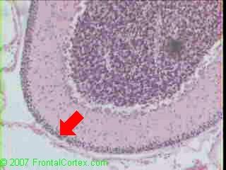
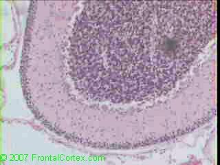
This slide shows neonatal cerebellum. Note the presence of the external granular cell layer (EGCL), which is indicated by the unnecessarily large red arrow in the image above. Cells from the EGCL migrate inward, to form the granule cell layer. The EGCL is seen in fetal and neonatal cerebellum.
(See References) | | |
| | |
| Please log in if you want to rate questions. | |||||
B) Adolescent cerebellum
This answer is incorrect.
This slide shows neonatal cerebellum. Note the presence of the external granular cell layer (EGCL), which is indicated by the unnecessarily large red arrow in the image above. Cells from the EGCL migrate inward, to form the granule cell layer. The EGCL is seen in fetal and neonatal cerebellum.
(See References) | | |
| | |
| Please log in if you want to rate questions. | |||||
C) This could be cerebellum from an adult or from an adolescent
This answer is incorrect.
This slide shows neonatal cerebellum. Note the presence of the external granular cell layer (EGCL), which is indicated by the unnecessarily large red arrow in the image above. Cells from the EGCL migrate inward, to form the granule cell layer. The EGCL is seen in fetal and neonatal cerebellum.
(See References) | | |
| | |
| Please log in if you want to rate questions. | |||||
D) This is choroid plexus
This answer is incorrect.
This slide shows neonatal cerebellum. Note the presence of the external granular cell layer (EGCL), which is indicated by the unnecessarily large red arrow in the image above. Cells from the EGCL migrate inward, to form the granule cell layer. The EGCL is seen in fetal and neonatal cerebellum.
(See References) | | |
| | |
| Please log in if you want to rate questions. | |||||
E) Neonatal cerebellum
This answer is correct.
This slide shows neonatal cerebellum. Note the presence of the external granular cell layer (EGCL), which is indicated by the unnecessarily large red arrow in the image above. Cells from the EGCL migrate inward, to form the granule cell layer. The EGCL is seen in fetal and neonatal cerebellum.
(See References) | | |
| | |
| Please log in if you want to rate questions. | |||||
References:
| 1. Prayson, R.A., and Goldblum, J.R. (Eds.) (2005). Neuropathology. Elsevier Churchill Livingstone, Philadelphia. |
| | |
| | |
| Please log in if you want to rate questions. | |||||
FrontalCortex.com -- Neurology Review Questions -- Neurology Boards -- Board Review -- Residency Inservice Training Exam -- RITE Exam Review
pathology
Cerebellar Microanatomy 01
Question ID: 112407099
Question written by J. Douglas Miles, (C) 2006-2009, all rights reserved.
Created: 11/24/2007
Modified: 11/24/2007
Estimated Permutations: 600
User Comments About This Question:
log in to FrontalCortex.com
New to FrontalCortex?
|

![]()
![]()

| | We comply with the HONcode standard for trustworthy health information: verify here. |
Share this page:
 |  |
 |
 |
 |
 |
 |
 |
 |
 |
 |
 |
 |
|
Saturday, July 05, 2025 at 12:50:00 PM
This site has been visited 49847919 times since June 6 2006
All software and content (C) 2004-2025, FrontalCortex, Inc. unless otherwise specified.
privacy policy Web 2.0 policy disclaimer contact us
All software and content (C) 2004-2025, FrontalCortex, Inc. unless otherwise specified.
privacy policy Web 2.0 policy disclaimer contact us