Vascular Malformations 04
Topic: Pathology
Created on Wednesday, November 28 2007 by jdmiles
Last modified on Wednesday, November 28 2007.
Which of the following statements about the type of lesion seen in this photomicrograph is most accurate?
This question was created on November 28, 2007 by jdmiles.
This question was last modified on November 28, 2007.
ANSWERS AND EXPLANATIONS
A) patients with this type of lesion have an initial presentation of focal epilepsy in 1/3 of cases
This answer is incorrect.
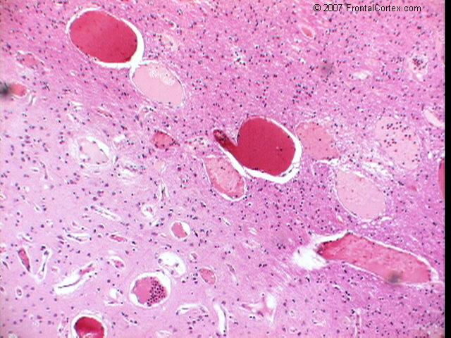
The photomicrograph shows a capillary telangiectasia. Capillary telangiectases are dilated capillaries, separated by normal brain tissue. They are usually incidental findings, and rarely become symptomatic. They have an estimated prevalence of 4 per 1000, comprising 16% to 20% of all brain vascular malformations. (
See References)
|
 |  |  | 
|  |  |
| Please log in if you want to rate questions. |
B) this type of lesion is associated with a 1% annual risk of acute hemorrhage
This answer is incorrect.
The photomicrograph shows a capillary telangiectasia. Capillary telangiectases are dilated capillaries, separated by normal brain tissue. They are usually incidental findings, and rarely become symptomatic. They have an estimated prevalence of 4 per 1000, comprising 16% to 20% of all brain vascular malformations. (
See References)
|
| | |
| | |
| Please log in if you want to rate questions. |
C) this is a capillary telangiectasia
This answer is correct.
The photomicrograph shows a capillary telangiectasia. Capillary telangiectases are dilated capillaries, separated by normal brain tissue. They are usually incidental findings, and rarely become symptomatic. They have an estimated prevalence of 4 per 1000, comprising 16% to 20% of all brain vascular malformations. (
See References)
|
| | |
| | |
| Please log in if you want to rate questions. |
D) lesions of this type are abnormal collections of thin-walled vessels without intervening brain tissue
This answer is incorrect.
The photomicrograph shows a capillary telangiectasia. Capillary telangiectases are dilated capillaries, separated by normal brain tissue. They are usually incidental findings, and rarely become symptomatic. They have an estimated prevalence of 4 per 1000, comprising 16% to 20% of all brain vascular malformations. (
See References)
|
| | |
| | |
| Please log in if you want to rate questions. |
E) this type of lesion accounts for 1.5%-4% of all brain masses
This answer is incorrect.
The photomicrograph shows a capillary telangiectasia. Capillary telangiectases are dilated capillaries, separated by normal brain tissue. They are usually incidental findings, and rarely become symptomatic. They have an estimated prevalence of 4 per 1000, comprising 16% to 20% of all brain vascular malformations. (
See References)
|
| | |
| | |
| Please log in if you want to rate questions. |
References:
| 1. Prayson, R.A., and Goldblum, J.R. (Eds.) (2005). Neuropathology. Elsevier Churchill Livingstone, Philadelphia. (ISBN:0443066582) | Advertising:
|
|
| | |
| | |
| Please log in if you want to rate questions. |
FrontalCortex.com -- Neurology Review Questions -- Neurology Boards -- Board Review -- Residency Inservice Training Exam -- RITE Exam Review
pathology
Vascular Malformations 04
Question ID: 11280703
Question written by J. Douglas Miles, (C) 2006-2009, all rights reserved.
Created: 11/28/2007
Modified: 11/28/2007
Estimated Permutations: 237600