Basic Neuropathology 05
Topic: Pathology
Created on Saturday, December 8 2007 by jdmiles
Last modified on Saturday, December 8 2007.
A) Necrosis
B) Amyloid plaques
C) Rosenthal fibers
D) Inflammatory demyelination
E) Granulomatous disease
 | = Go back to the top of the page. |
 | = Reload a different version of this question (). |
 | = Load a random question from the database. |

| = Use this question as a template to create a totally NEW question. |
 | = Enter detailed rating for this question! |
 | = How users like you have rated this question. |
This question was last modified on December 08, 2007.
ANSWERS AND EXPLANATIONS
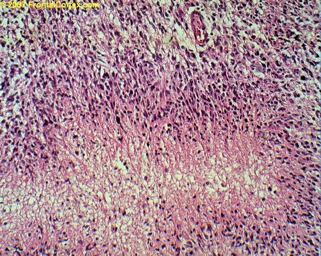
A) Necrosis
This answer is correct.
The abnormalities we are looking at in this slide are pseudopallisading and necrosis. "Pallisading" is when cells line up next to each other, like slats in a picket fence. These cells appear to pallisade around the central pale area, which represents necrotic tissue. Pseudopallisading and necrosis are findings associated with glioblastoma multiforma (GBM), which is the tumor shown in this slide. (See References)
| | |
| | |
| Please log in if you want to rate questions. | |||||
B) Amyloid plaques
This answer is incorrect.
The abnormalities we are looking at in this slide are pseudopallisading and necrosis. "Pallisading" is when cells line up next to each other, like slats in a picket fence. These cells appear to pallisade around the central pale area, which represents necrotic tissue. Pseudopallisading and necrosis are findings associated with glioblastoma multiforma (GBM), which is the tumor shown in this slide. (See References)
| | |
| | |
| Please log in if you want to rate questions. | |||||
C) Rosenthal fibers
This answer is incorrect.
The abnormalities we are looking at in this slide are pseudopallisading and necrosis. "Pallisading" is when cells line up next to each other, like slats in a picket fence. These cells appear to pallisade around the central pale area, which represents necrotic tissue. Pseudopallisading and necrosis are findings associated with glioblastoma multiforma (GBM), which is the tumor shown in this slide. (See References)
| | |
| | |
| Please log in if you want to rate questions. | |||||
D) Inflammatory demyelination
This answer is incorrect.
The abnormalities we are looking at in this slide are pseudopallisading and necrosis. "Pallisading" is when cells line up next to each other, like slats in a picket fence. These cells appear to pallisade around the central pale area, which represents necrotic tissue. Pseudopallisading and necrosis are findings associated with glioblastoma multiforma (GBM), which is the tumor shown in this slide. (See References)
| | |
| | |
| Please log in if you want to rate questions. | |||||
E) Granulomatous disease
This answer is incorrect.
The abnormalities we are looking at in this slide are pseudopallisading and necrosis. "Pallisading" is when cells line up next to each other, like slats in a picket fence. These cells appear to pallisade around the central pale area, which represents necrotic tissue. Pseudopallisading and necrosis are findings associated with glioblastoma multiforma (GBM), which is the tumor shown in this slide. (See References)
| | |
| | |
| Please log in if you want to rate questions. | |||||
References:
| 1. Prayson, R.A., and Goldblum, J.R. (Eds.) (2005). Neuropathology. Elsevier Churchill Livingstone, Philadelphia. (ISBN:0443066582) | Advertising: |
| | |
| | |
| Please log in if you want to rate questions. | |||||
FrontalCortex.com -- Neurology Review Questions -- Neurology Boards -- Board Review -- Residency Inservice Training Exam -- RITE Exam Review
pathology
Basic Neuropathology 05
Question ID: 120807148
Question written by J. Douglas Miles, (C) 2006-2009, all rights reserved.
Created: 12/08/2007
Modified: 12/08/2007
Estimated Permutations: 60480
User Comments About This Question:
log in to FrontalCortex.com
New to FrontalCortex?
|

![]()
![]()

| | We comply with the HONcode standard for trustworthy health information: verify here. |
Share this page:
 |  |
 |
 |
 |
 |
 |
 |
 |
 |
 |
 |
 |
|
Monday, June 30, 2025 at 5:38:04 PM
This site has been visited 49812232 times since June 6 2006
All software and content (C) 2004-2025, FrontalCortex, Inc. unless otherwise specified.
privacy policy Web 2.0 policy disclaimer contact us
All software and content (C) 2004-2025, FrontalCortex, Inc. unless otherwise specified.
privacy policy Web 2.0 policy disclaimer contact us