Muscle Fiber Histology 01
Topic: Pathology
Created on Thursday, December 20 2007 by jdmiles
Last modified on Thursday, December 20 2007.
A) Denervated muscle fibers appear darker
B) Type 2 fibers normally stain darker than type 1 fibers
C) Muscle fibers affected by Duchenne muscular dystrophy appear darker
D) Type 1 fibers normally stain darker than type 2 fibers
E) Type 1 fibers normally stain lighter than type 2 fibers
 | = Go back to the top of the page. |
 | = Reload a different version of this question (). |
 | = Load a random question from the database. |

| = Use this question as a template to create a totally NEW question. |
 | = Enter detailed rating for this question! |
 | = How users like you have rated this question. |
This question was last modified on December 20, 2007.
ANSWERS AND EXPLANATIONS
A) denervated muscle fibers appear darker
This answer is incorrect.
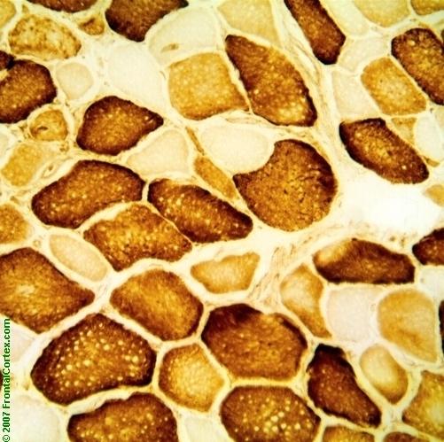
Cytochrome C oxidase is a marker of mitochondrial activity. Type 1 muscle fibers have a higher capacity for oxidative metabolism than type 2 cells, which have a higher capacity for anaerobic metabolism. This means that the type 1 muscle cells will stain darker. (See References)
| | |
| | |
| Please log in if you want to rate questions. | |||||
B) type 2 fibers normally stain darker than type 1 fibers
This answer is incorrect.
Cytochrome C oxidase is a marker of mitochondrial activity. Type 1 muscle fibers have a higher capacity for oxidative metabolism than type 2 cells, which have a higher capacity for anaerobic metabolism. This means that the type 1 muscle cells will stain darker. (See References)
| | |
| | |
| Please log in if you want to rate questions. | |||||
C) muscle fibers affected by Duchenne muscular dystrophy appear darker
This answer is incorrect.
Cytochrome C oxidase is a marker of mitochondrial activity. Type 1 muscle fibers have a higher capacity for oxidative metabolism than type 2 cells, which have a higher capacity for anaerobic metabolism. This means that the type 1 muscle cells will stain darker. (See References)
| | |
| | |
| Please log in if you want to rate questions. | |||||
D) type 1 fibers normally stain darker than type 2 fibers
This answer is correct.
Cytochrome C oxidase is a marker of mitochondrial activity. Type 1 muscle fibers have a higher capacity for oxidative metabolism than type 2 cells, which have a higher capacity for anaerobic metabolism. This means that the type 1 muscle cells will stain darker. (See References)
| | |
| | |
| Please log in if you want to rate questions. | |||||
E) type 1 fibers normally stain lighter than type 2 fibers
This answer is incorrect.
Cytochrome C oxidase is a marker of mitochondrial activity. Type 1 muscle fibers have a higher capacity for oxidative metabolism than type 2 cells, which have a higher capacity for anaerobic metabolism. This means that the type 1 muscle cells will stain darker. (See References)
| | |
| | |
| Please log in if you want to rate questions. | |||||
References:
| 1. Prayson, R.A., and Goldblum, J.R. (Eds.) (2005). Neuropathology. Elsevier Churchill Livingstone, Philadelphia. (ISBN:0443066582) | Advertising: |
| | |
| | |
| Please log in if you want to rate questions. | |||||
FrontalCortex.com -- Neurology Review Questions -- Neurology Boards -- Board Review -- Residency Inservice Training Exam -- RITE Exam Review
pathology
Muscle Fiber Histology 01
Question ID: 122007034
Question written by J. Douglas Miles, (C) 2006-2009, all rights reserved.
Created: 12/20/2007
Modified: 12/20/2007
Estimated Permutations: 8400
User Comments About This Question:
log in to FrontalCortex.com
New to FrontalCortex?
|

![]()
![]()

| | We comply with the HONcode standard for trustworthy health information: verify here. |
Share this page:
 |  |
 |
 |
 |
 |
 |
 |
 |
 |
 |
 |
 |
|
Monday, June 30, 2025 at 5:30:36 PM
This site has been visited 49812153 times since June 6 2006
All software and content (C) 2004-2025, FrontalCortex, Inc. unless otherwise specified.
privacy policy Web 2.0 policy disclaimer contact us
All software and content (C) 2004-2025, FrontalCortex, Inc. unless otherwise specified.
privacy policy Web 2.0 policy disclaimer contact us